April 25, 2024
New 3D X-ray microscope supports orthopaedic research at Waterloo
Equipment allows researchers to see inside tissues and medical devices
Equipment allows researchers to see inside tissues and medical devices
One in eight Canadians experiences bone and joint dysfunction due to aging, injury and disease, and a new 3D X-ray microscope at the University of Waterloo will support new orthopaedic research and development to improve surgical treatment of musculoskeletal conditions.
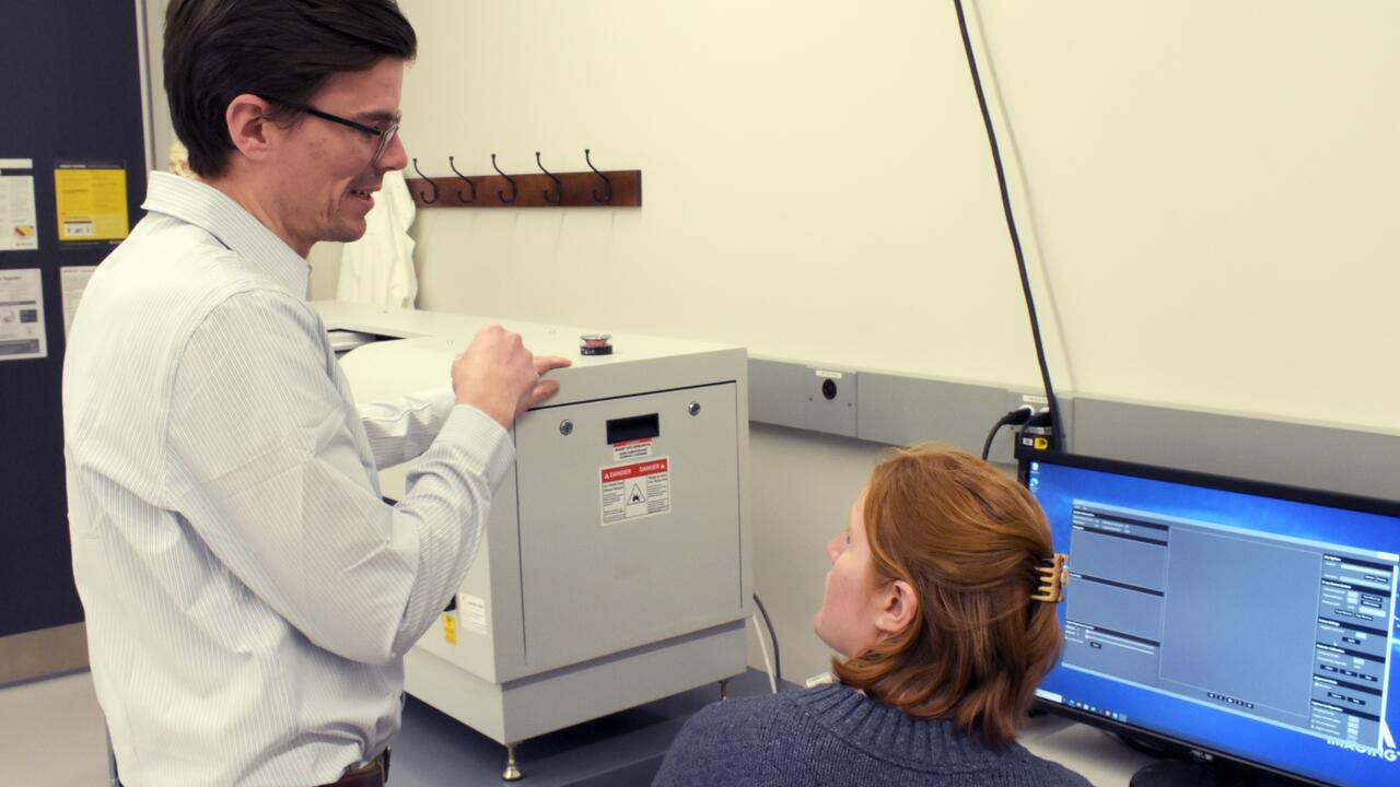
The inCiTe™ 3D X-ray Microscope will be used in projects led by Dr. Stewart McLachlin, a professor in the Department of Mechanical and Mechatronics Engineering at Waterloo, who conducts biomedical research as part of the Orthopaedic Mechatronics Laboratory.
"This is very exciting as this equipment allows us to see inside biological tissues and medical devices to understand how the internal microstructure is formed and deformed under varied conditions,” said McLachlin, who leads the new lab with Dr. Naveen Chandrashekar.
According to McLachlin, Waterloo is the first research facility in Canada to have access to this phase-contrast 3D X-ray microscope imaging for biological tissue characterization.
“The phase-contrast technology improves the image contrast and resolution of the internal microstructure for lower density materials like bone and joint tissues, overcoming a major limitation of traditional CT imaging,” McLachlin said. “This will allow us to better understand tissue mechanics, and in turn, work towards developing better orthopaedic implants and treatment methods.”
The inCiTe™ 3D X-ray Microscope was developed by KA Imaging. It is the first commercial system that utilizes BrillianSe™, a patented high-spatial resolution amorphous selenium (a-Se) detector exclusively developed by KA Imaging Inc. Dr. Karim Karim, a professor in the Department of Electrical and Computer Engineering at Waterloo, is the company’s chief technology officer.
The NSERC Research Tools and Instruments grant program supported the purchase of this system.