In 2011, Jane Holbrook and Mary Power from the Centre for Teaching Excellence organized a workshop for large class instructors: "Enjoying Your Large Class (Making the Most of Your Large Class)". An activity in this session was to "design a 'blended' activity (one that students participate in online and in class) to promote student engagement in a specific course topic". From this workshop and activity, the Microbiology Art Gallery was born.
The Microbiology Art Gallery allows students in my Fundamentals of Microbiology class to explore course content artistically. No stress, no marks, just art. The annual gallery has invited creative students to enrich the class (and themselves) with their talents. Announced during the 2nd week of term, the art gallery remains open all term for submissions on the course website. Just before class, after a submission is received, I dim the lights for us to admire the artwork as a group. Usually I offer comments about context and the medium used to prepare, sometimes I ask the student a question, and the class joins me in applauding the artist. At the end of the term we use our clickers to vote on "audience favourites" and gift bags are awarded.
Even though participation is voluntary, every year there are 20-30 submissions (i.e., 2-3% participation) that provide unexpected, impressive, thought-provoking, quirky, and often beautiful perspectives on course content. I quickly discovered that large university science classes are a goldmine for untapped creative talent - our students are amazing. Below are a few examples of submissions I've received over the years with brief comments about how they relate to course content. Note that when students uploaded their work, they agree to the use of their contribution for department/course purposes provided that their name is attributed.
Lauren Singroy (2011)
This colourful painting captures a bacterial virus ("bacteriophage") - the mustache is a tip of the hat to our class' participation in Movember activities.
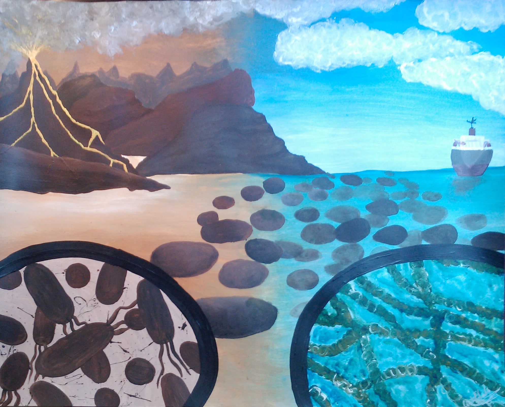
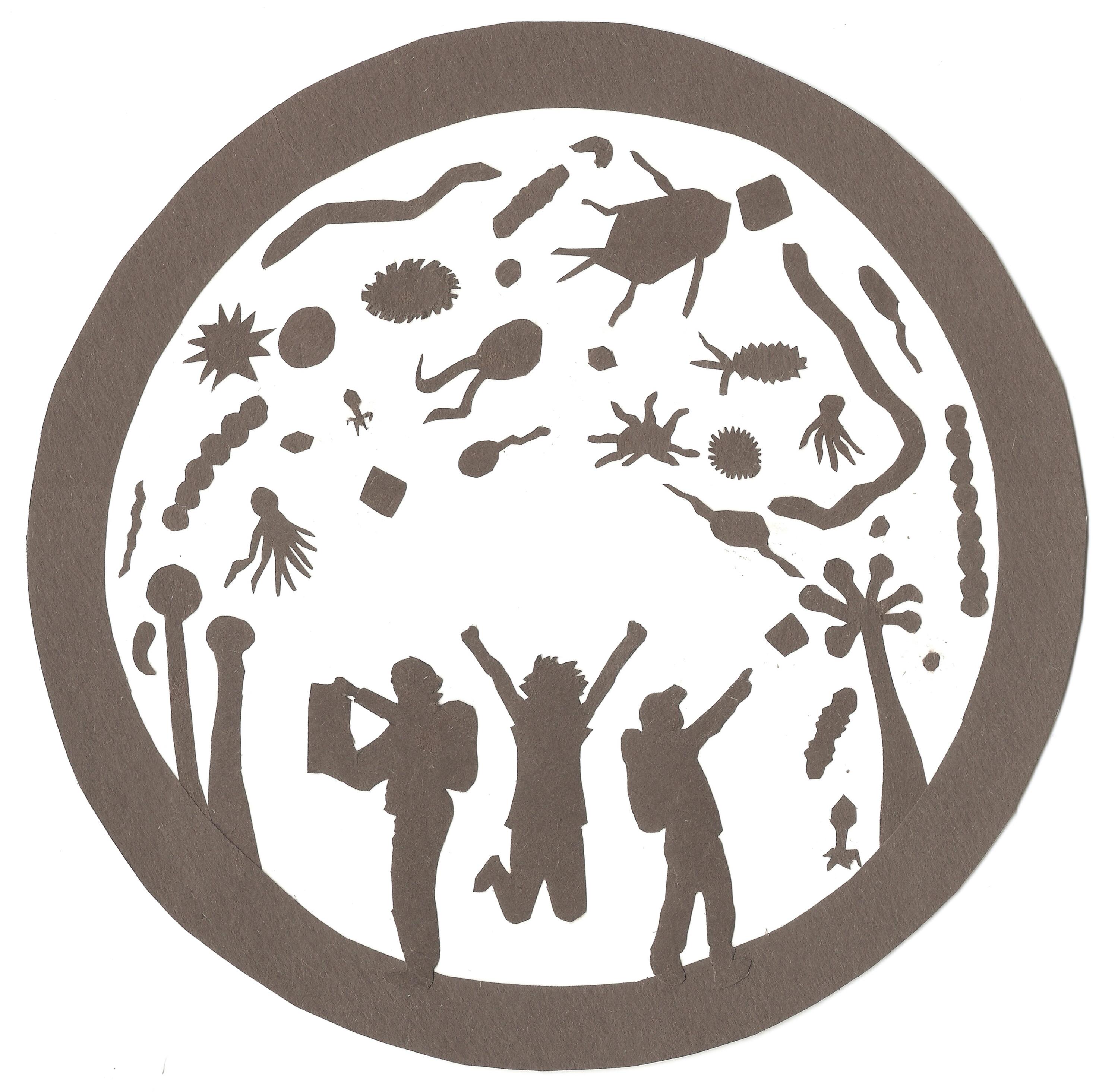
Chitrita Chakko (2011)
I love how Chitrita captured the microbiological timeline of Earth with her painting, from mineral eating microbes early on (left side) to stromatolite pedestals (center) that hosted the earliest forms of O2-producing photosynthetic cyanobacteria (right). Present day, with boat, at the right.
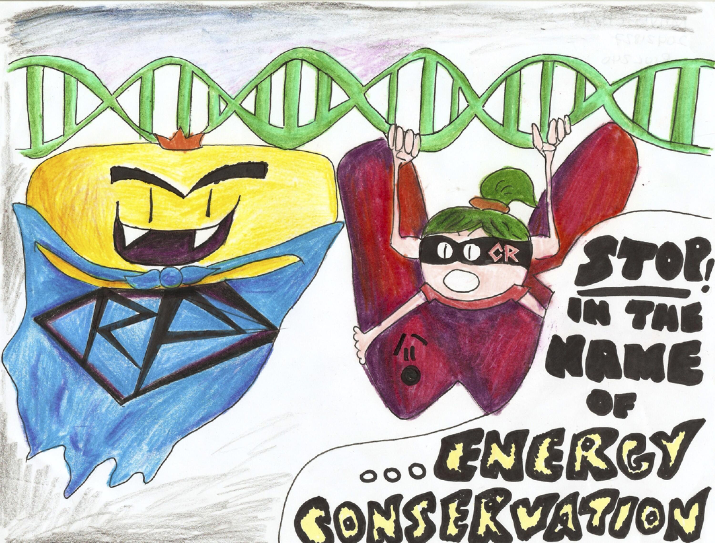
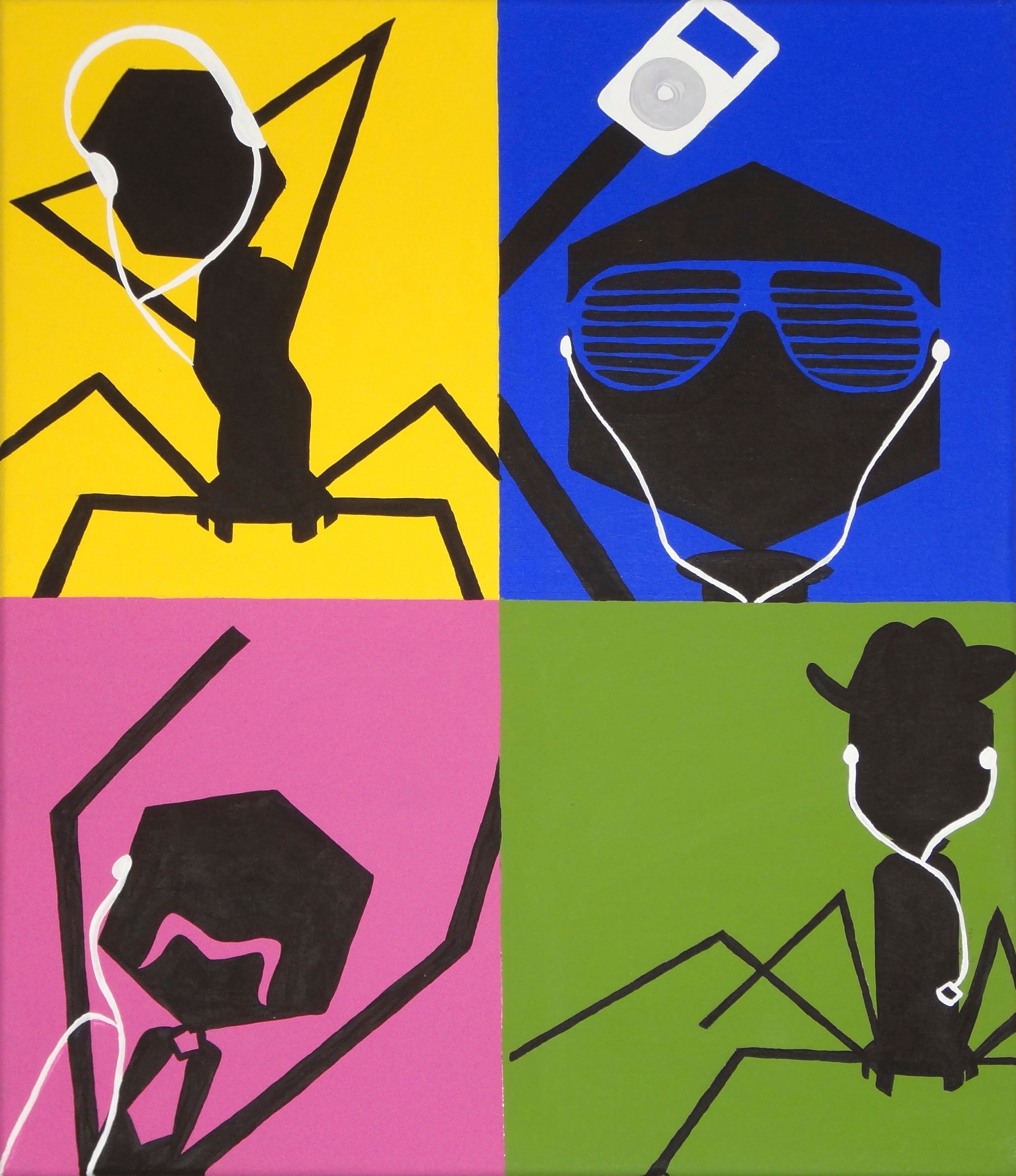
Mavis Chan (2012)
Regulation of gene expression is very important for a cell in its efforts to conserve energy. This can happen by RNA polymerase (yellow) being unable to convert DNA to RNA because a repressor protein (red) is blocking the polymerase's path because of a co-repressor (with mask). Superhero themed - love it!
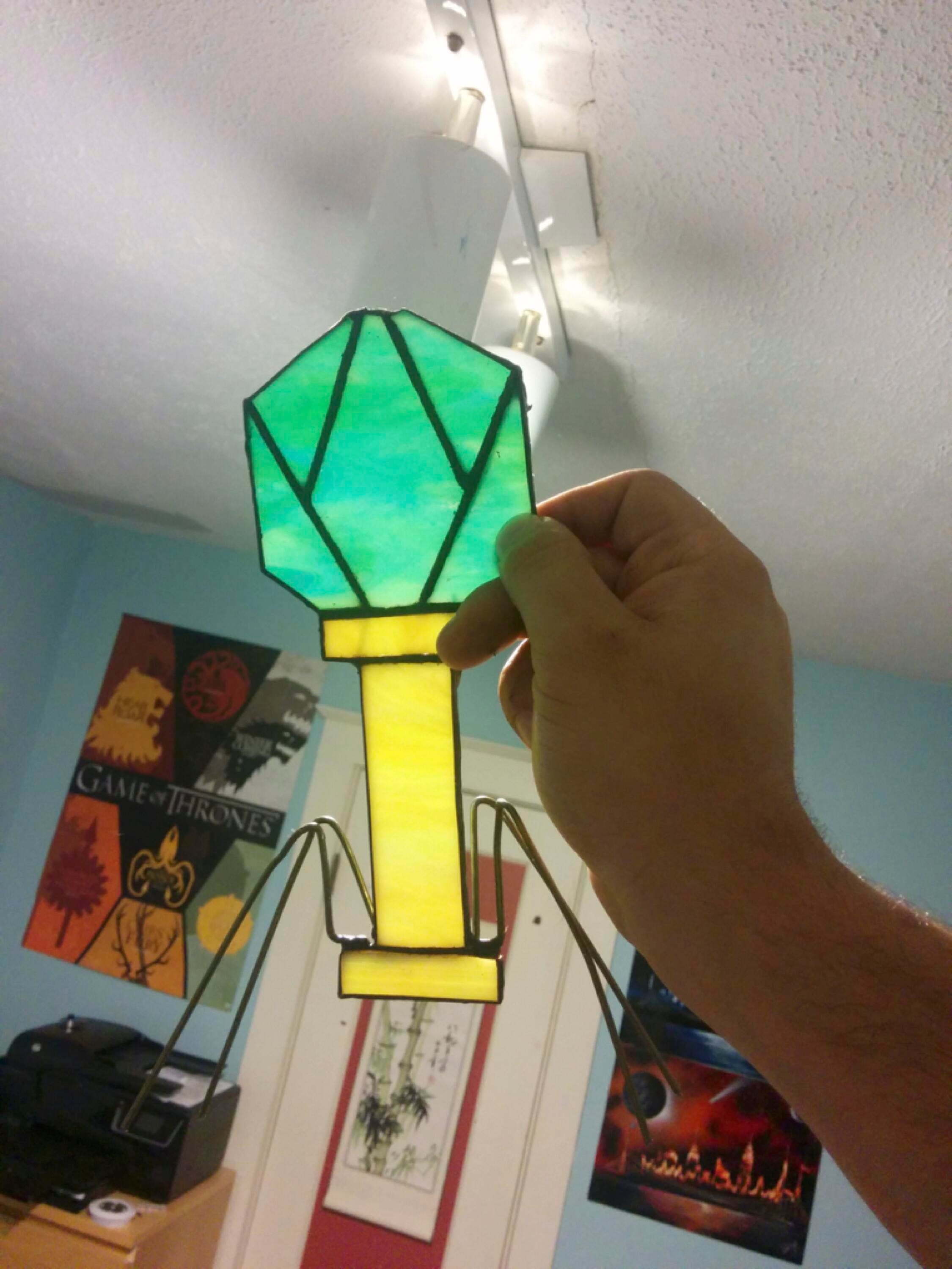
Mark Zanon (2014)
The remarkable geometric shapes associated with bacterial viruses capture student imaginations each year, especially the syringe-like structure of E. coli-infecting bacteriophage T4. I agree with Mark that stained glass is a fitting medium to reproduce this virus shape.
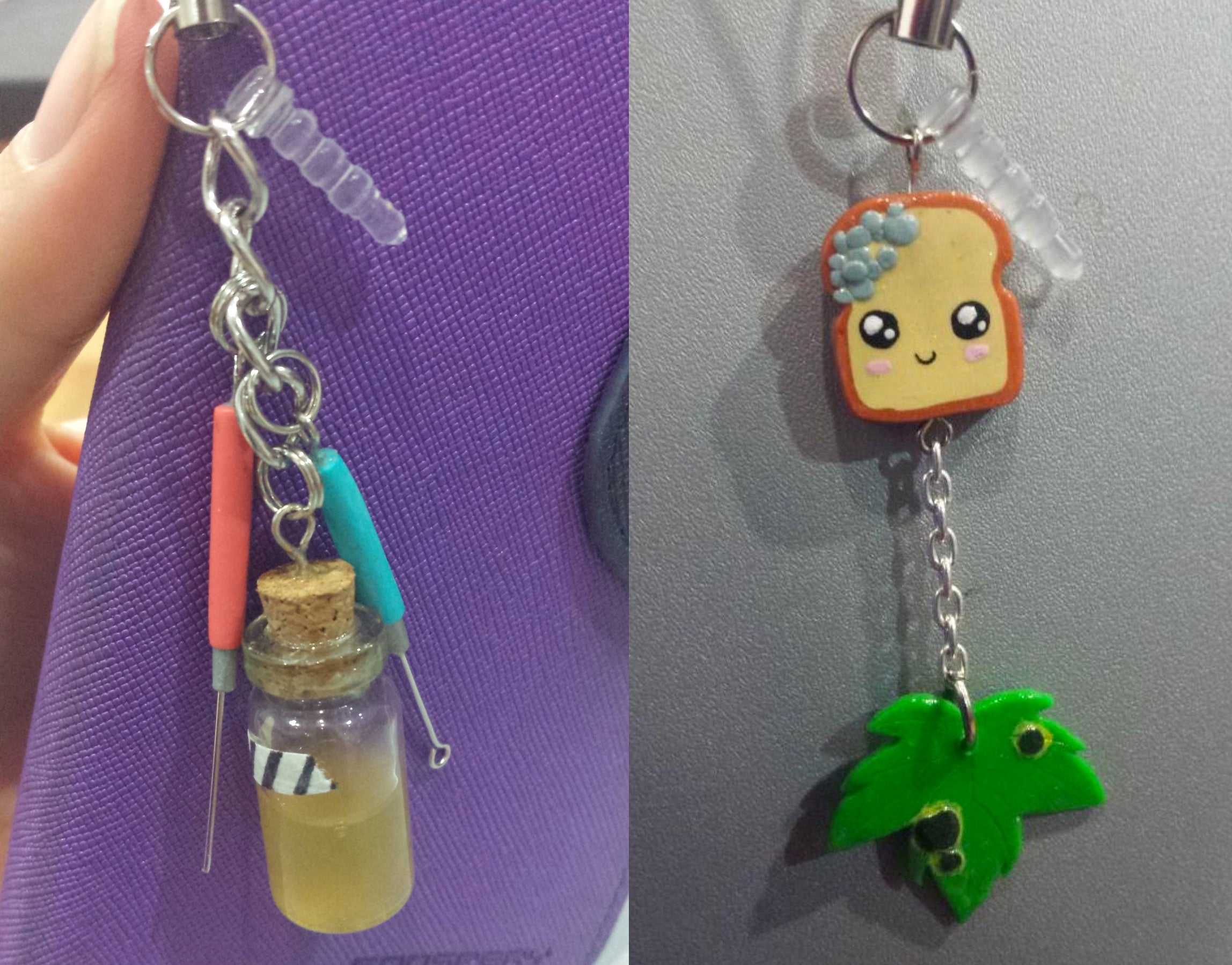
Cindy Luu (2014)
These were the first dangle phone charms I'd seen - they hang from phone audio plugs - and they are surprisingly lifelike! On the left is a stab (pink handle) and loop (turqoise handle) for inoculating culture deeps and broths/plates, respectively. The autoclave tape is a nice touch! On the right are fungal infections of bread (Penicillium) and maple leaves (Rhytisma).
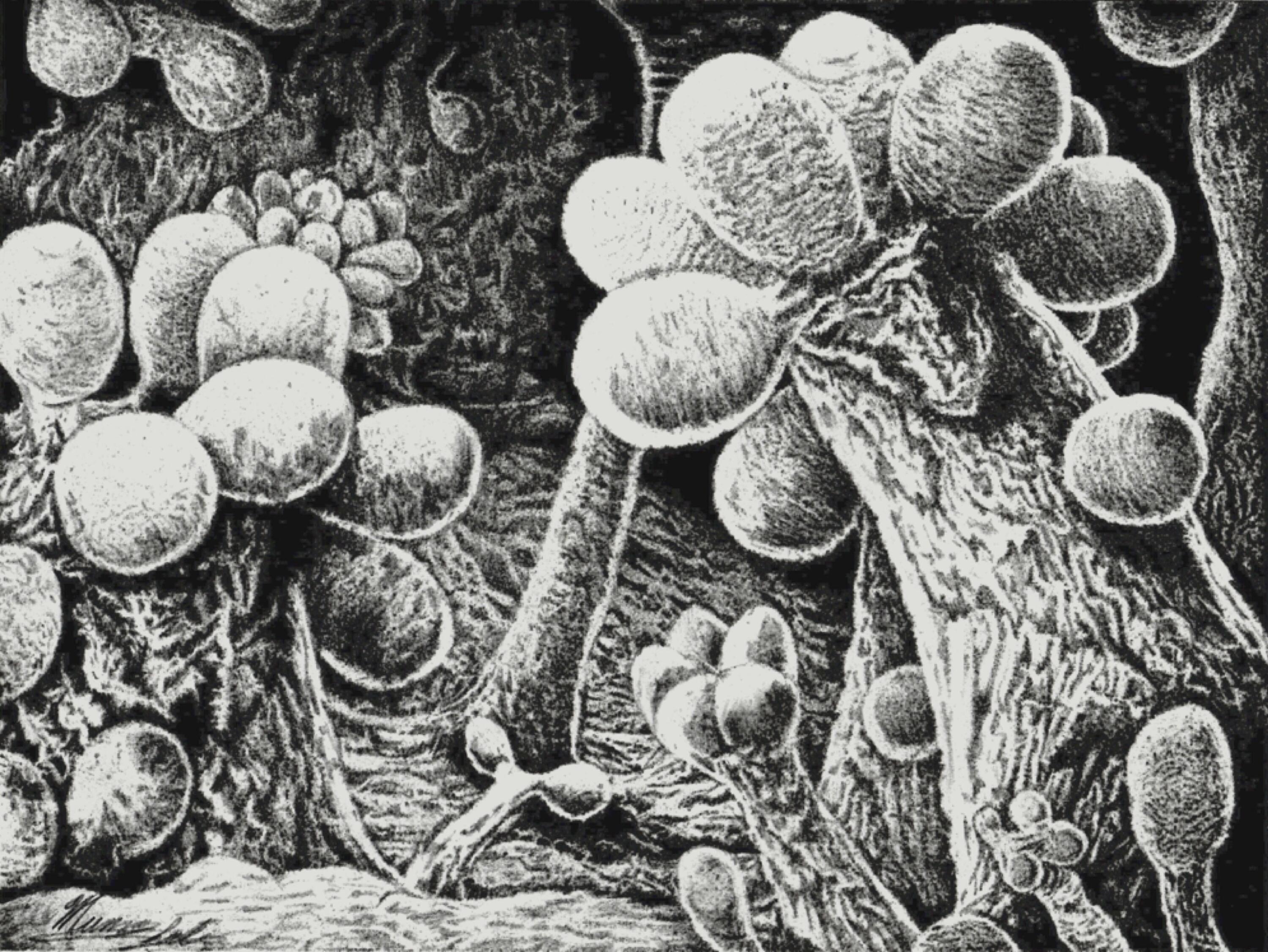
Munaza Saleem (2014)
Addmittedly, I make a big deal in class about how remarkable myxobacteria are as predatory soil bacteria. They swarm, hunt, communicate, and aggregate into millimeter high visible structures that differentiate to send their spores into hungry passing snails for deposit elsewhere. Amazing bacteria, as is Munaza's pencil drawing of their fruiting bodies.
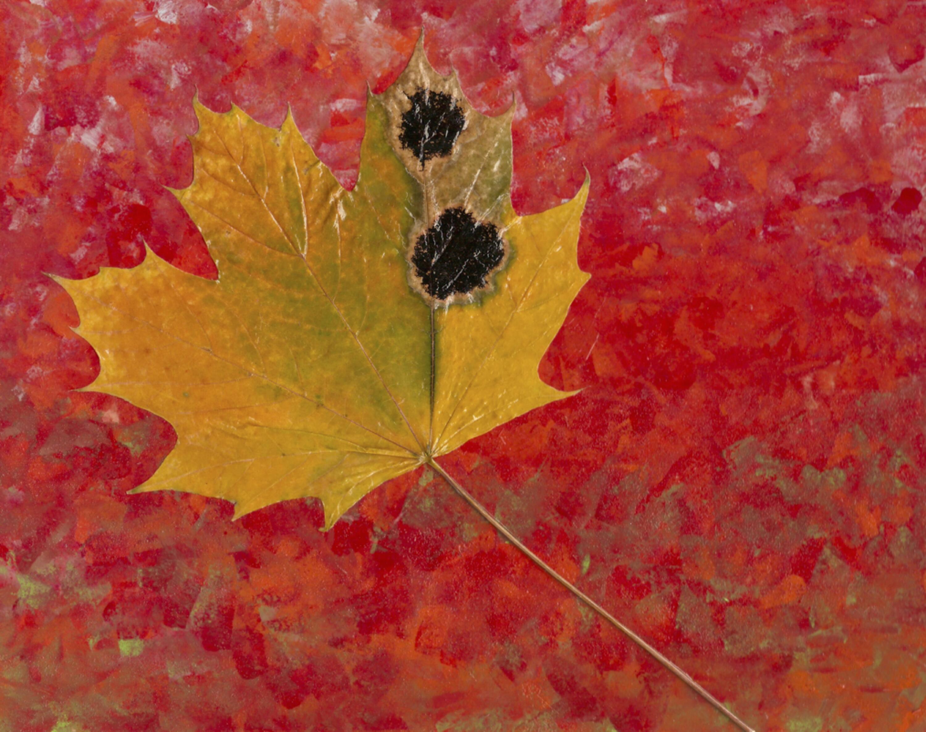
Rachel Beaver (2014)
Every year I send students out to find Rhytisma and have them post #rhytisma selfies on social media to "confuse the internet". Often I'll bring bags of infected leaves with me to class, encouraging students to get "up close and personal" with these fungal parasites of maple trees that cause no harm to humans. This leaf on canvas version is a uniquely permanent way to portray the infection.
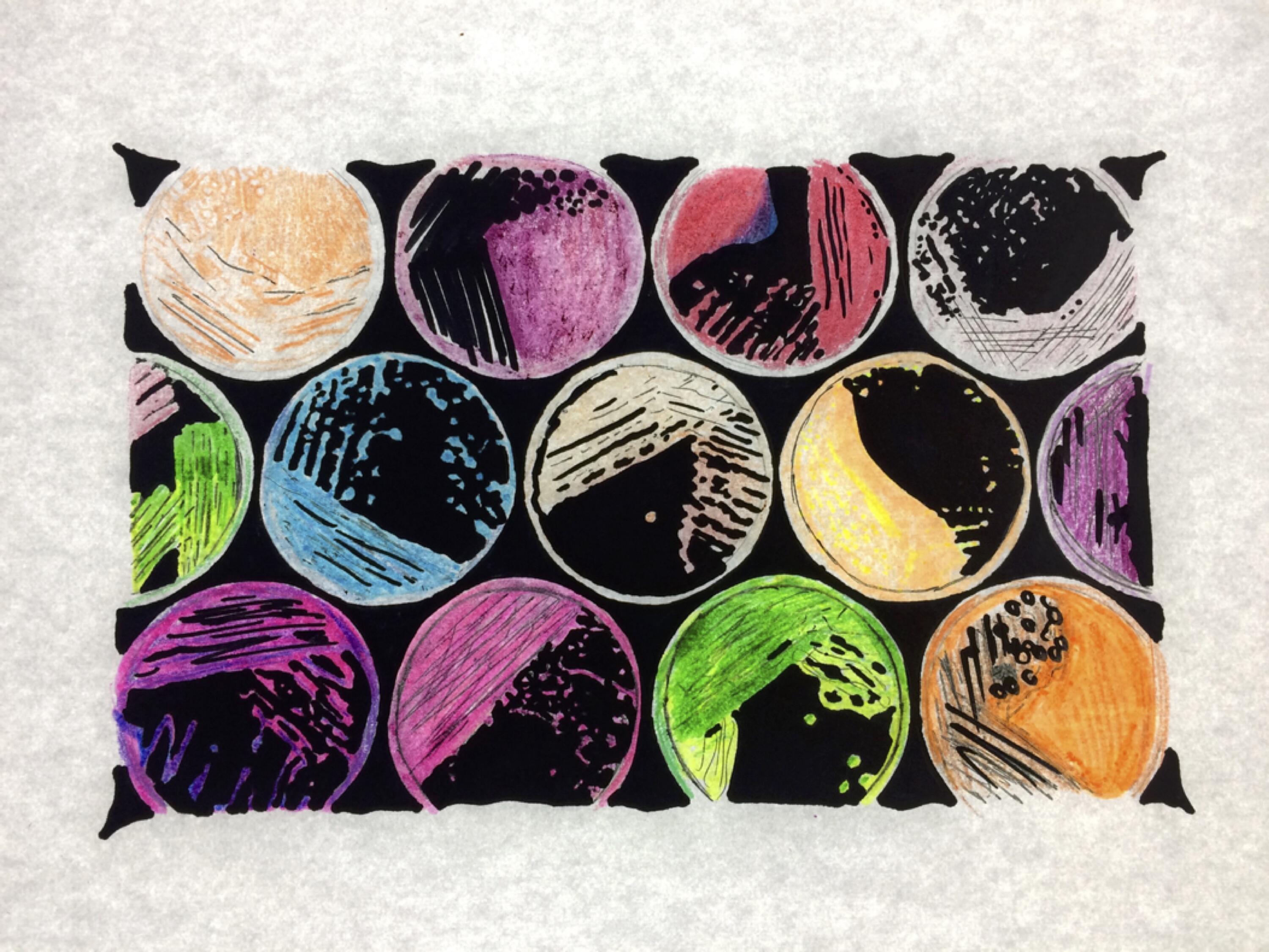
Emma Duarte (2017)
Petri plates with streaks of bacteria - love the colours!
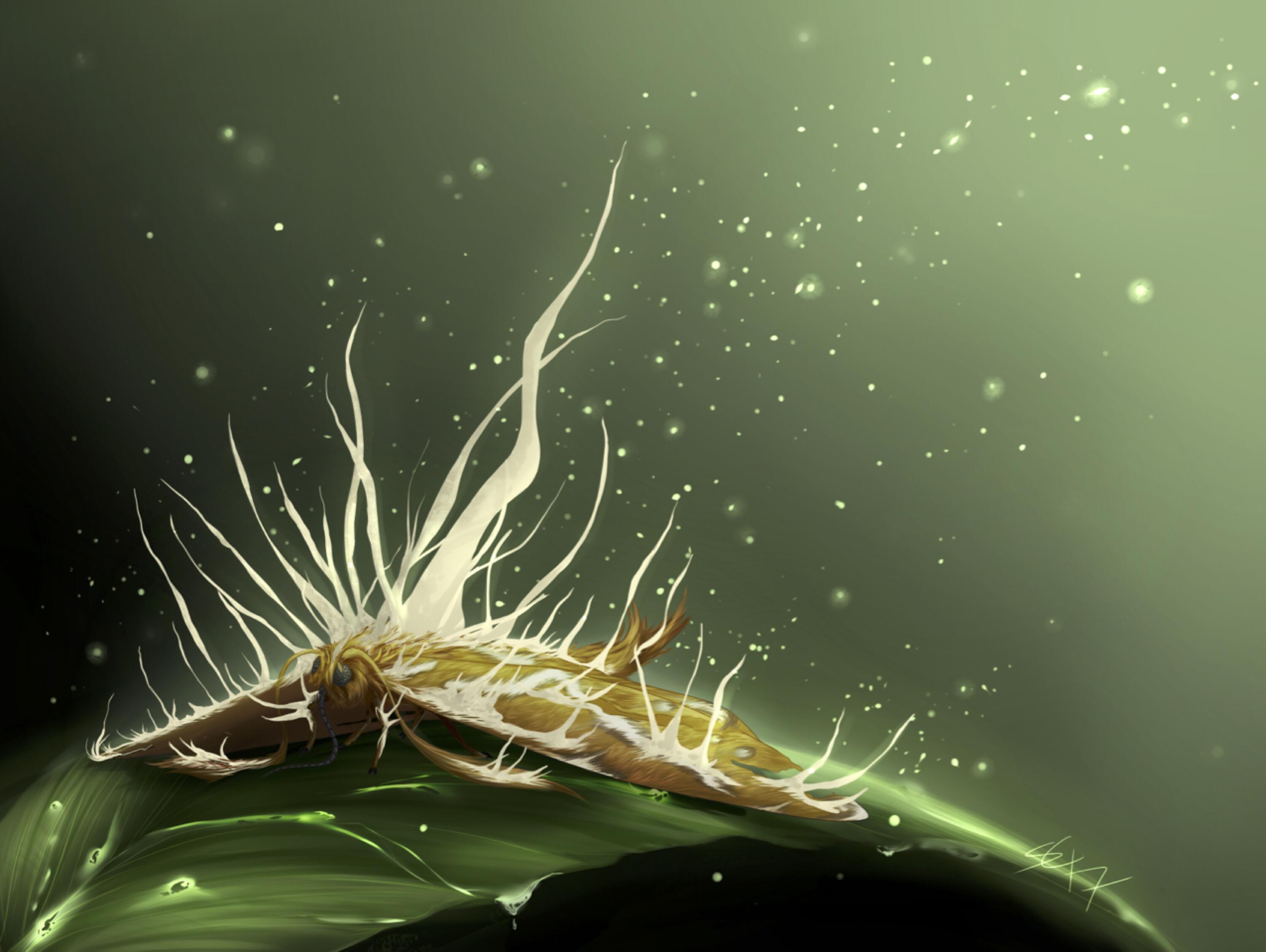
Sarah Cook (2017)
The ability of Cordyceps fungi to infect insects, control their behaviours, and then produce fruiting bodies from their corpses amazes and disgusts students every year. Many are aware that ants can be infected (from the Planet Earth Attenborough documentary) but there are many species of Cordyceps adapted to many different arthropods, as shown by this striking artistic rendition of Cordyceps mushrooms from a moth.
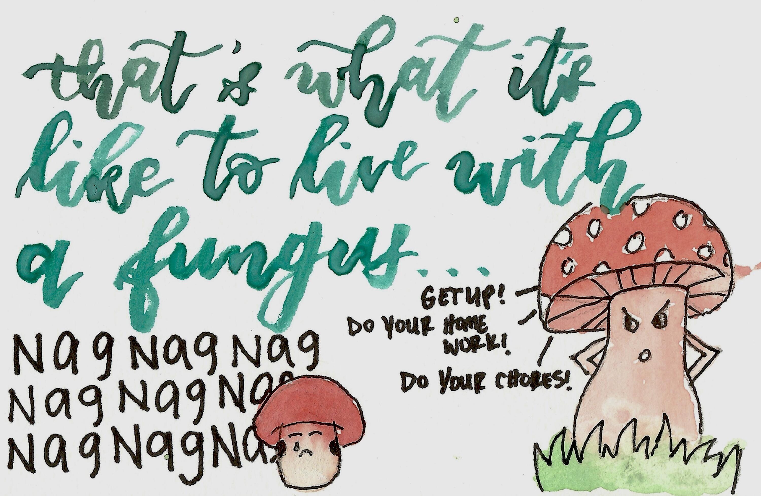
Seanthel Santos (2017)
When explaining how fungi have cell walls of chitin, I explain that chitin is simply a polymer of N-acetylglucosamine (NAG) - so NAG NAG NAG is what it's like to live with a fungi (I've been heard to say apparently). Like Seanthel, many students find ways of expressing humour through course content - lots of cartoon submissions over the years :)
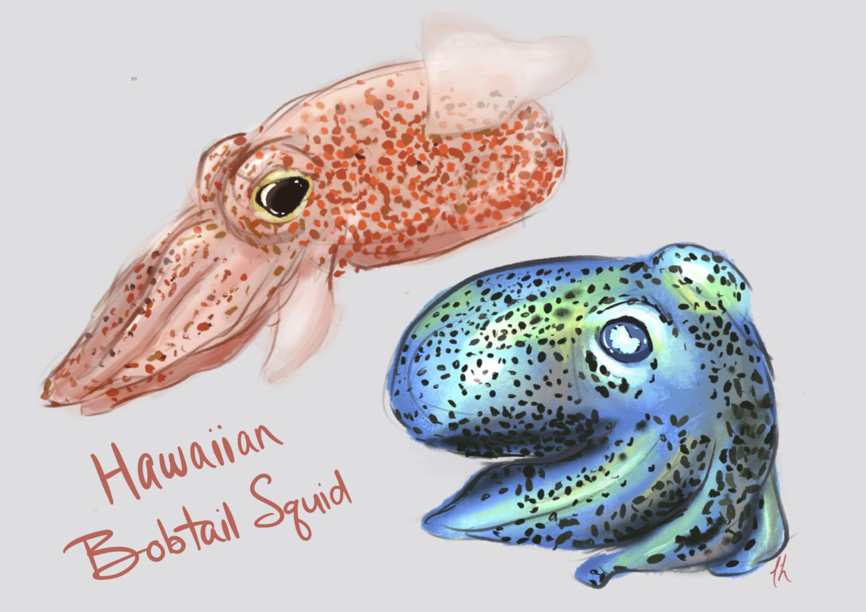
Lisa Hopkins (2018)
Students are fascinated by the way certain bacteria that produce light inhabit the "light organ" of the Hawaiin bobtail squids, which helps squid avoid casting a shadow when out and about at night. I can go on and on about the bacteria and these squid but for now it's good enough to indicate that some of the most remarkable gallery submissions (like this one) focus on these photogenic cephalopods.
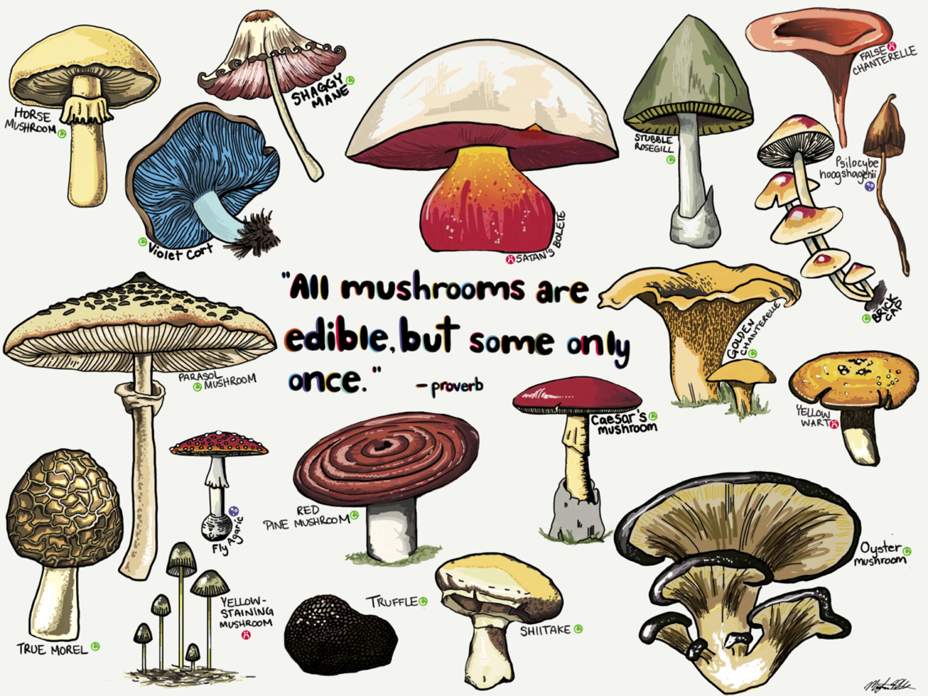
Meghan Paleolog (2018)
My enthusiasm for mushrooms shines through the course and is picked up by students like Meghan who beautifully captured some of the most (in)famous specimens, along with one of my favourite quotes to share with the class each year.
Jacob Contini (2019)
The flavour of art gallery submissions is often responsive to current events, such as the release of Bohemian Rhapsody in 2018. This creative contribution reminds me of an LP I have somewhere on a shelf in the basement...
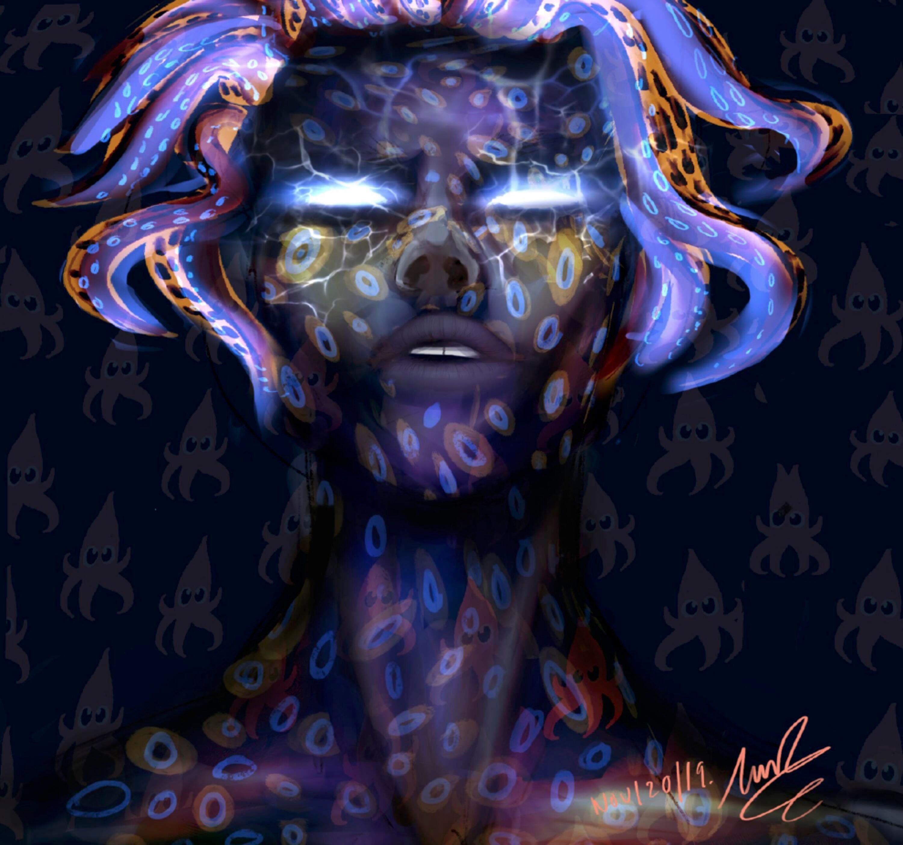
Tianna Rodrigues (2019)
Look closely - see the hair and background squid? Another contribution that celebrates the Hawaiin bobtail squid and its light-producing bacteria. This example was submitted with a time-lapse video showing the creative process itself, which I set to music for added classroom impact.
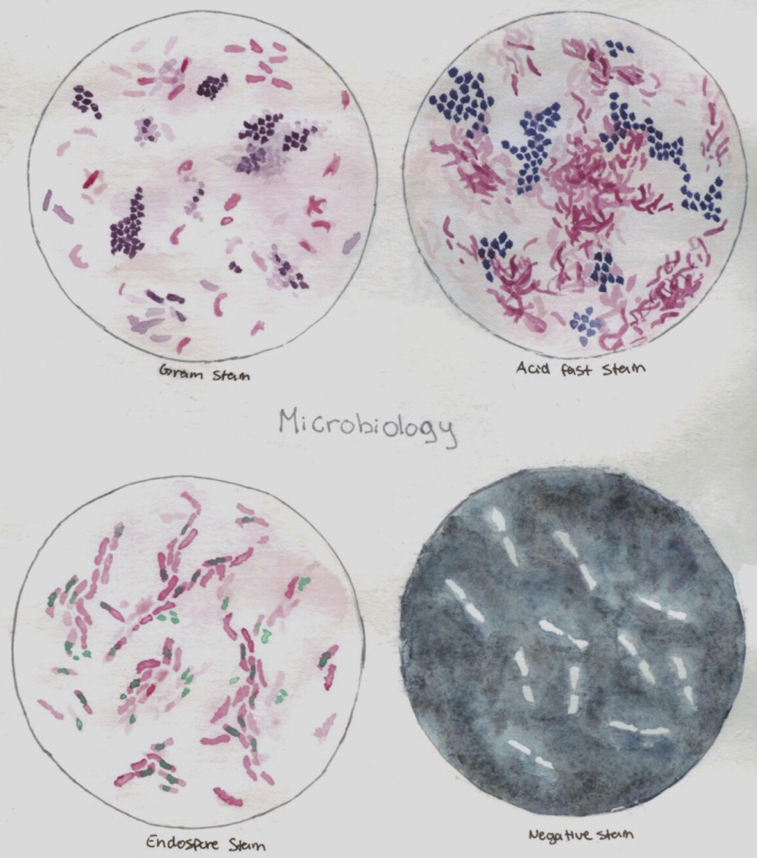
Olivia Li (2019)
Microscopy and stains are a centrepiece of undergraduate microbiology labs, and this lovely watercolour nicely captures anticipated results for these classic stains.
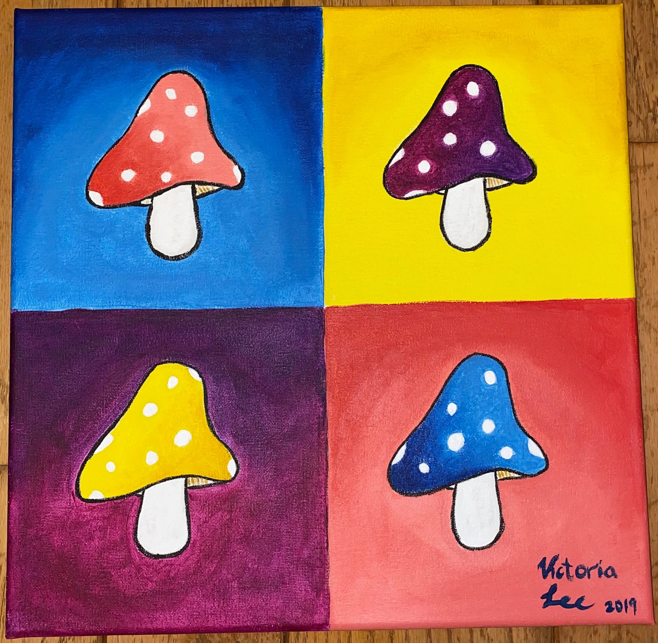
Victoria Lee (2019)
The 2011 bacteriophage painting from Lauren Singroy (that hangs on my office wall) inspired Victoria to create this similar collage during one of her office hour visits. Can't go wrong with fly agarics! (unless you eat them)
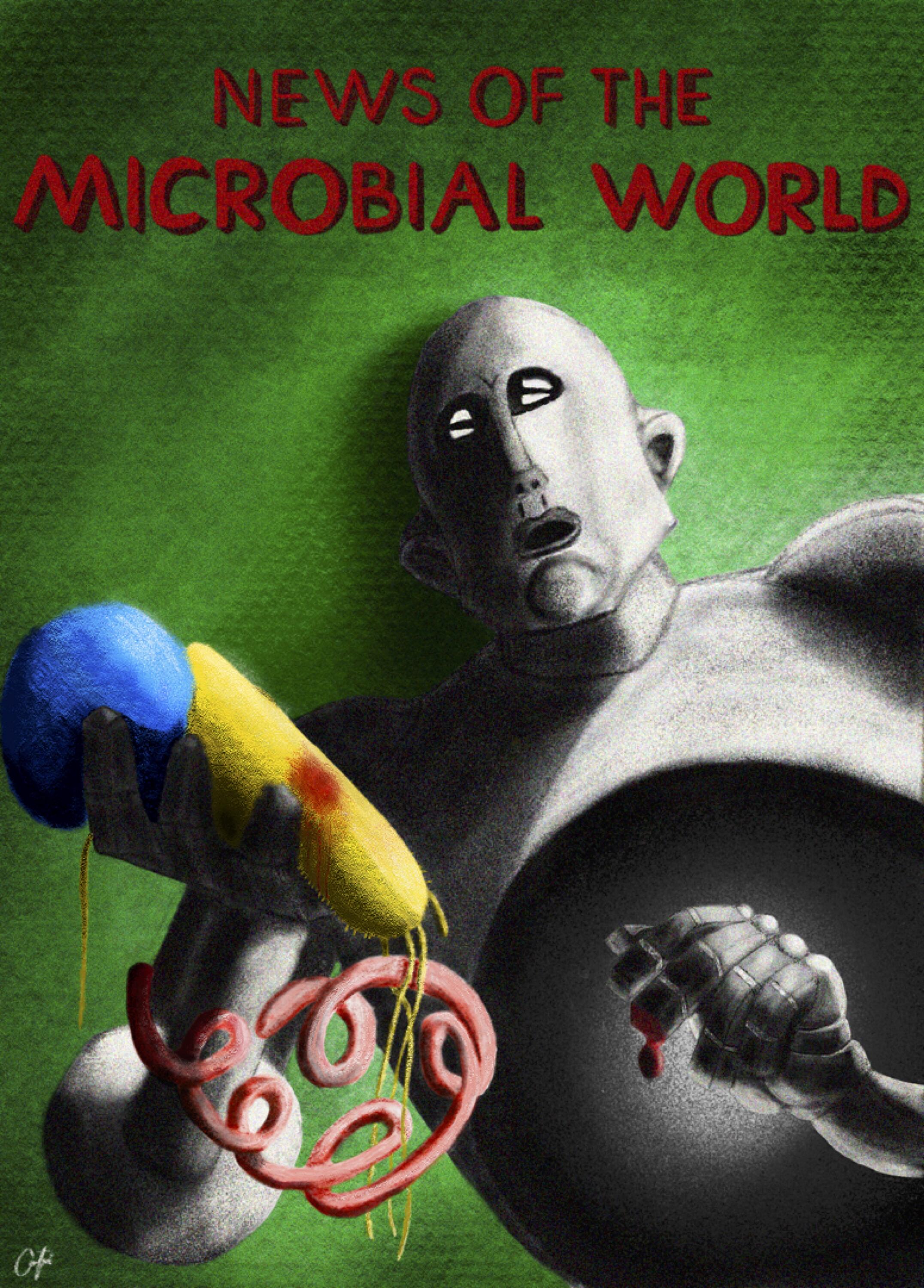
Mary Zach (2019)
This contribution captures the core of what teaching Fundamentals of Microbiology means to me. This course is my yearly opportunity to inspire hundreds of science students to fall in love with microbiology by exploring the magic of the microbial world. Thanks for capturing some of this excitement in your art gallery contribution Mary!