University of Waterloo researchers have invented a digital medical imaging system that significantly improves the cancer detection process to deliver immediate results and enable swift, effective treatment for all types of cancer.
The Photon Absorption Remote Sensing (PARS) system, an innovative, built-from-scratch technology, is faster than traditional cancer-detection methods and aims to deliver a diagnosis in minutes — enabling prompt surgical intervention. Currently, patients can wait weeks or even months to receive biopsy results, leading to delays in treatment and increased patient anxiety.
The new system is also highly accurate. Clinical studies using human breast tissue showed that pathologists were unable to distinguish between images generated by the PARS system and those produced by conventional methods. The technology boasts a 98 per cent correlation with traditional diagnostic techniques.
"Our primary goal is to provide patients with timely and accurate diagnoses, reducing the need for multiple surgeries and minimizing the risk of cancer spread," said Dr. Parsin Haji Reza, lead researcher and a professor in Waterloo’s Department of Systems Design Engineering.
“This invention will transform digital pathology, enabling surgeons to obtain multiple results simultaneously with just one biopsy and provide accurate diagnoses within minutes. It also ensures thorough removal of cancerous tissue before closing the incision, mitigating the need for further surgeries.”
The biopsy process is a crucial step in determining the presence of cancer. The traditional histopathology method involves taking a tissue sample and preparing it for analysis — cutting it into sections, staining those sections with different dyes, mounting those sections onto slides, and analyzing those slides under a microscope to identify cancer markers.
By replacing traditional methods with high-resolution imaging powered by artificial intelligence (AI), the PARS system accelerates the diagnostic process, saving both time and resources. Lasers are applied to a tissue sample which generates a unique and detailed set of data in high resolution. The data is fed into the AI system which translates it into a standard histopathology image for the pathologist to read. It removes the need for multiple slide preparation by applying digitized image filters to the one tissue sample providing multiple reads — all without damaging the sample which allows for further analysis if needed.
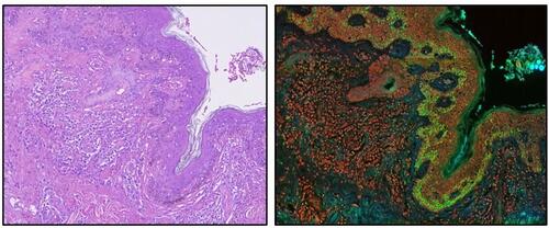
Comparison of traditional histopathology imaging vs. PARS raw data
“In addition to reducing patients’ stress, this technology will save billions of dollars for the health care system,” said Reza. “Surgeon and pathologists’ time costs money, each slide and special dye costs money, the hospitals’ facilities cost money. We can now reduce all those costs with faster biopsy results that are just as accurate.”
The research is supported by grants from the Natural Sciences and Engineering Research Council of Canada (NSERC) and the Canadian Institutes of Health Research (CIHR) and has garnered significant interest from investors and pharmaceutical companies.
illumiSonics, a Waterloo-based startup, has been established to commercialize the research with plans to bring the technology to market within two years. The company offers graduate students involved in the research employment opportunities to continue to steer the system’s development.
More information about this work can be found in the recent research papers Automated Whole Slide Imaging for Label-Free Histology using Photon Absorption Remote Sensing Microscopy, published in IEEE Transactions on Biomedical Engineering, and Multi-channel feature extraction for virtual histological staining of photon absorption remote sensing images, published in Scientific Reports.
This article was originally posted on the Waterloo News website.
Check out the CTV news article for more details