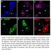
The Skeletal Muscle Biology & Cell Death Laboratory is equipped with state-of-the-art equipment and uses the latest cellular and molecular approaches to study skeletal muscle signaling, structure, function, phenotype, and viability. We work with a variety of experimental models including primary cell cultures and cell lines, normal and transgenic mice, healthy and diseased rodents, and humans.
Cell Culture Facilities
The Skeletal Muscle Biology & Cell Death Laboratory has access to its own dedicated cell culture room equipped with cell culture hoods, CO2 incubators, inverted microscope, and automated cell size/count analyzer. Scroll through image gallery below for more information.
Imaging Cytometry, Flow Cytometry & Cell Sorting
The Skeletal Muscle Biology & Cell Death Laboratory has an in-house multicolor flow cytometer and cell sorter, as well as a multi-color imaging cytometer. This equipment is ideal for various types of cell analyses, cell sorting, organelle assays (mitochondria), cytokine analysis (multiplex beads), and high throughput imaging of various cellular structures and events. Scroll through image gallery below for more information.
Microscopy
The Skeletal Muscle Biology & Cell Death Laboratory has an in-house microscopy suite equipped with brightfield and fluorescent microscopes providing immunofluorescent (widefield, structured illumination, laser confocal) and immunohistochemical imaging. In addition, live-cell imaging is available through a stage-top incubation system. Sample preparation (cryostat) and staining equipment are also available. Scroll through image gallery below for more information.
High-Resolution Respirometry, Cell Imaging, Protein & Gene Analysis
The Skeletal Muscle Biology & Cell Death Laboratory is equipped with numerous additional pieces of equipment that allow for a variety of analyses and experiments to be conducted. Some of the equipment available including, high-resolution respirometer, plate cell imager, gel documentation systems, HPLC systems, real-time PCR systems, a nanodrop spectrophotometer, thermocyclers, plate spectrophotometer, plate spectrofluorometer, a muscle biophysics system, animal surgical equipment, and rodent treadmills. Scroll through image gallery below for more information.
Image Gallery


