What tools are we using to study cardiovascular physiology?
Conventional ultrasound
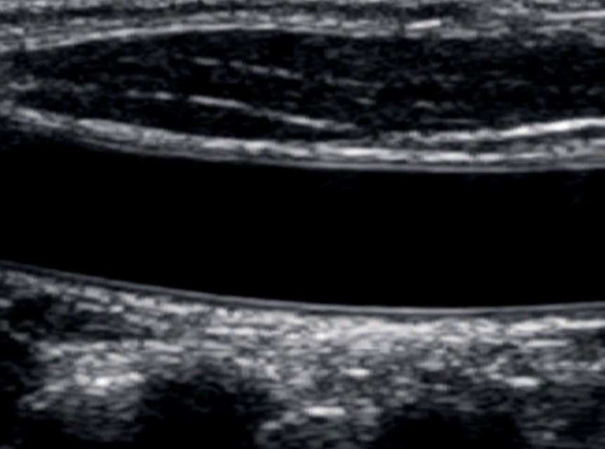
Pictured: A B-mode image of the common carotid artery, with clearly visible arterial layers on the near and far walls.
Non-invasive ultrasound imaging allows us to measure both the structure and function of the vascular system in humans with minimal risk. In the Vascular Observations through Research on Technology and Exercise (VORTEX) Lab, we use conventional ultrasound machines that are found in clinical environments to make basic observations about conduit arteries, the heart, and superficial veins. B-mode ultrasound allows us to view the thickness and kinematics of arterial walls across the cardiac cycle to learn more about how arterial structure changes with aging and disease processes. Doppler ultrasound allows us to measure the speed and flow of blood through vessels, which changes with activity and sendentary behaviour, and can tell us how blood is being supplied to end-organs.
Next-generation ultrasound
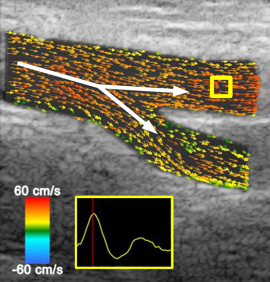
Pictured: A still vector flow image of the femoral bifurcation during peak blood flow, with smaller arrows indicating the direction and magnitude of individual pixels over time, and larger arrows indicating the general flow of blood through the bifurcation.
We are one of the only human physiology labs in the world to use open-platform high frame rate ultrasound (HiFRUS) to study cardiovascular function. As opposed to conventional ultrasound, HiFRUS platforms allow access to raw radiofrequency information to make customizable and highly-specific measurements about vascular function at >1000 fps. In the VORTEX Lab, we specialize in using vector flow imaging to visualize and quantify 'complex blood flow' found in special vascular regions such as arterial bifurcations and venous valves. Blood flow in these areas is not straight and creates unique eddies, jets, and, yes, vortexes, which are thought to be intricately linked to vessel function and disease risk. Taking advantage of this technology allows us to make discoveries in difficult-to-study areas of the circulatory system that have primary roles in the development of cardiovascular disease across the lifespan.
Pressure waveforms
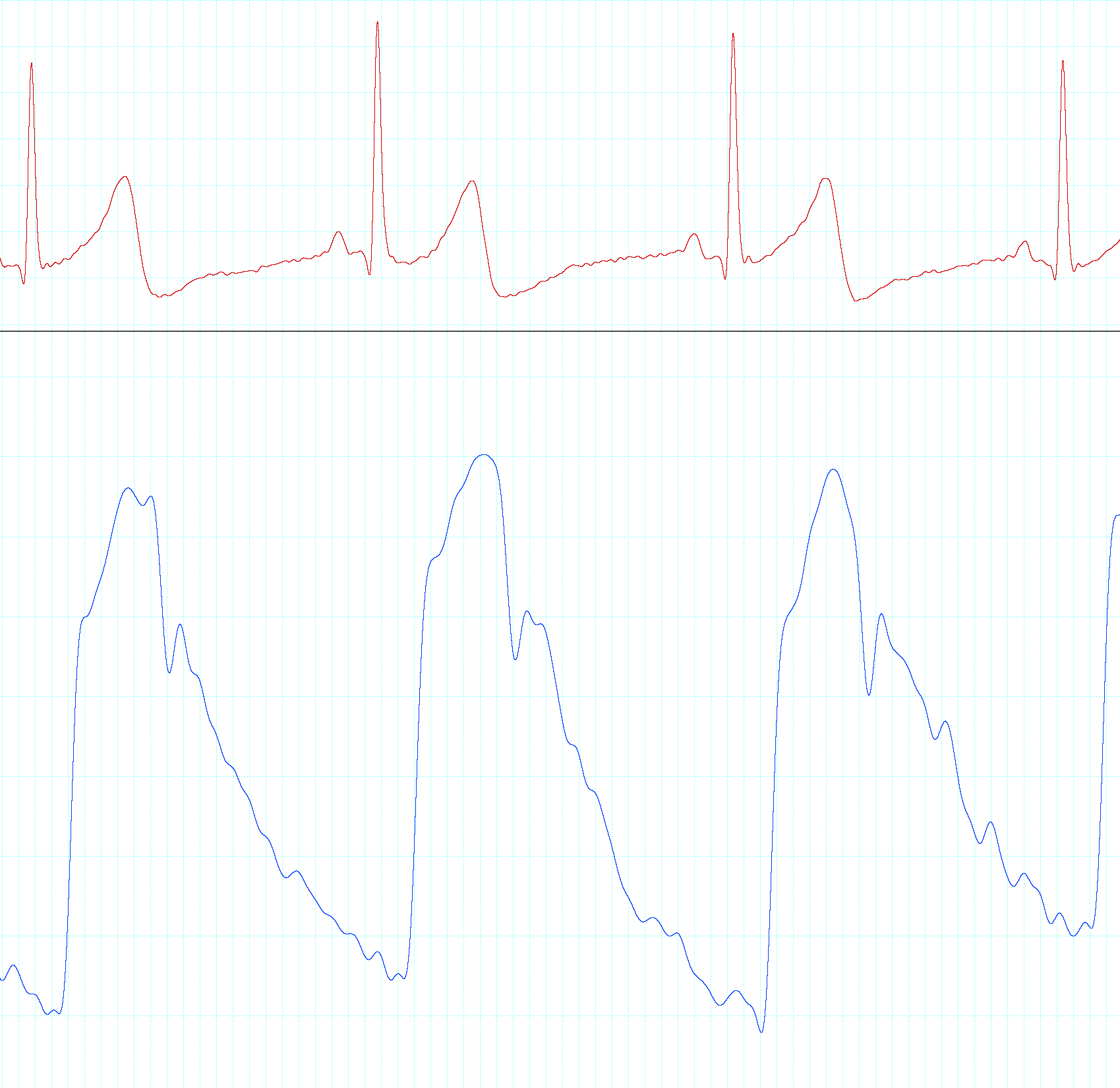
Pictured: A single-lead electrocardiogram trace above a pressure waveform of a 'young' carotid artery.
We use applanation tonometry to assess pressure waveforms in the major arteries of the body. These devices allow sensitive recordings of arterial pressure when arteries 'push' against pressure-sensitive pads each heart beat. We use these pressure waveforms to measure different properties of arteries such as arterial stiffness and arterial distensibility, which are known to predict the risk of cardiovascular disease later in life.