1 / 20
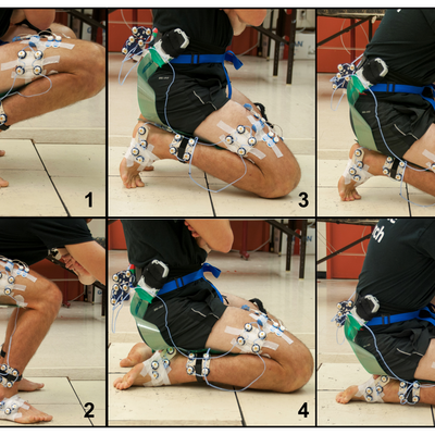
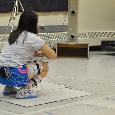
Sagittal view of six high knee flexion postures. 1) heels-up squat, 2) flatfoot squat, 3) dorsiflexed kneel, 4) plantarflexed kneel, 5) dorsiflexed asymmetric kneel, and 6) plantarflexed asymmetric kneel.

2 / 20
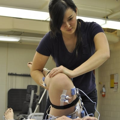
Performing passive range of motion for the hip joint (hip scour) of a participant prior to data collection to normalize across individual flexibility differences.

3 / 20
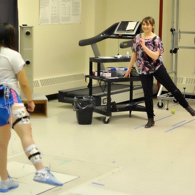
Footswitches allow us to identify the instant when points of a shoe or foot make contact with the ground during movement.

4 / 20
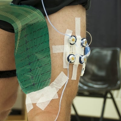
Attachment of a pressure sensor on a participant to measure force transfer between the thigh and shank segments during high knee flexion movements.

5 / 20
Functional trials allow us to identify hard to locate joint centers to improve our measurement of segment movement and force transmission through the body.

6 / 20
By strapping in multiple locations along different segments we can apply fixed weights to see how far the leg will abduct or adduct.
7 / 20
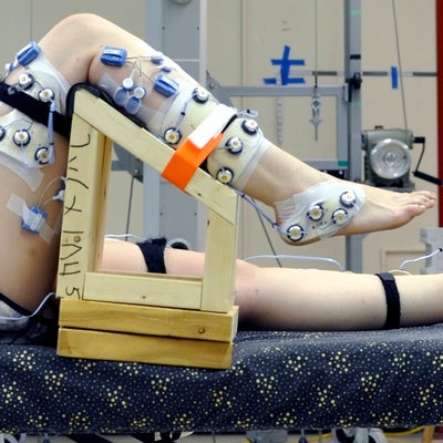
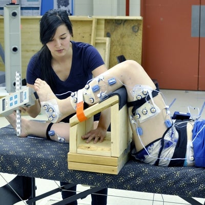
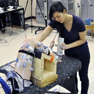
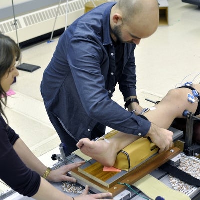
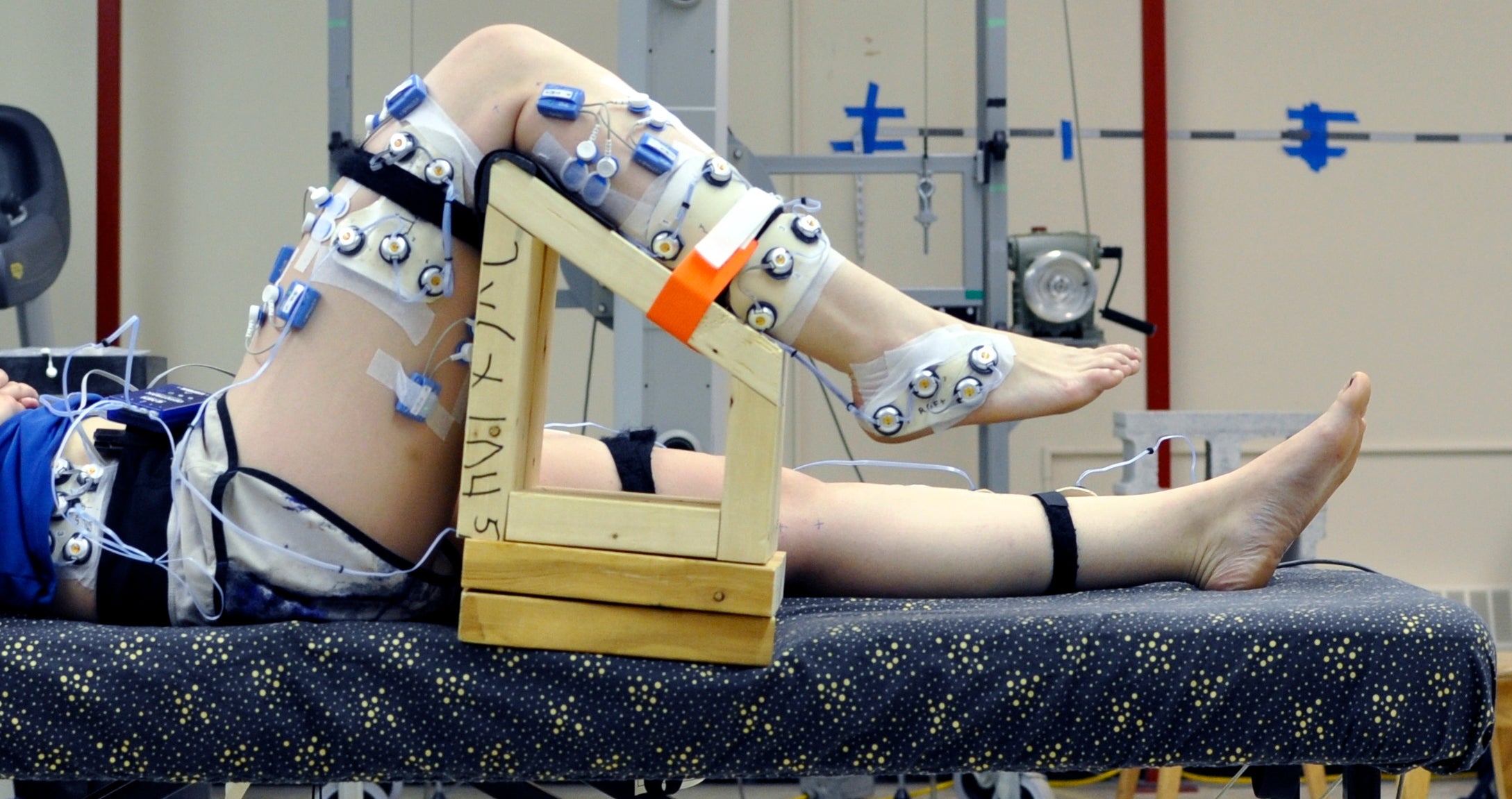
To test ankle range of motion we can support the participant's leg in a fixed position and apply forces to the foot.

8 / 20
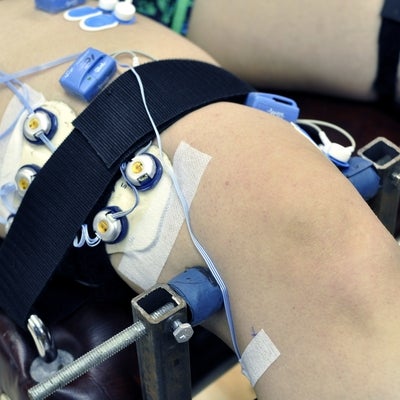
Femoral epicondyles are clamped onto a base with the thigh strapped down to keep it static during weight application to the shank.

9 / 20
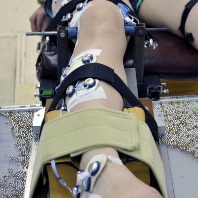
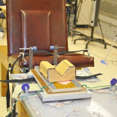
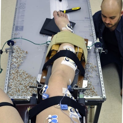
A padded sled rests atop ball bearings on a lexan table top, creating a nearly frictionless surface for measuring frontal plane knee joint laxity. The participant is seated in the chair and the leg is strapped into the padded cradle.

10 / 20
Applying a load to the sled to abduct the participant's leg in a safe and controlled manner.

11 / 20
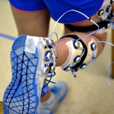
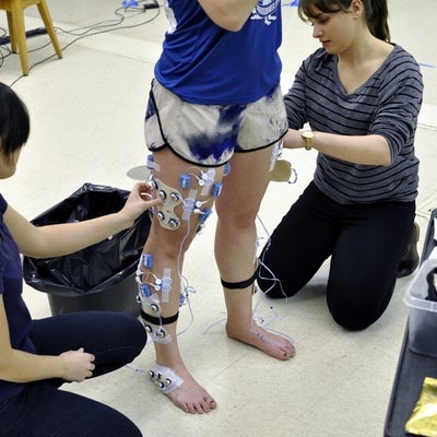
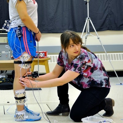
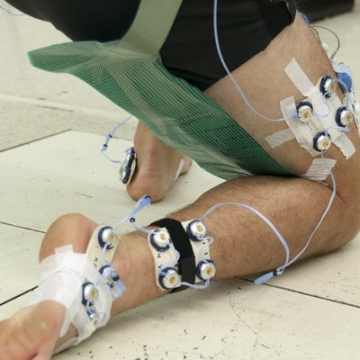
Instrumenting a participant's lower limb for motion tracking and electromyography data collection.

12 / 20
Placing a digitizing probe (Optotrak, Northern Digital Inc., Waterloo, ON) on a participant's knee to create a virtual landmark for 3D segment reconstruction.

13 / 20
Applying a measured force to maximally dorsiflex the participant's ankle to characterize this participant's ankle flexibility.

14 / 20
Applying a measured force to maximally plantarflex the participant's ankle to characterize this participant's ankle flexibility.

15 / 20
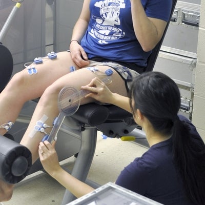
Measuring a participant's knee flexion angle prior to a maximum voluntary contraction to assure that their knee extensors can maximally activate.

16 / 20
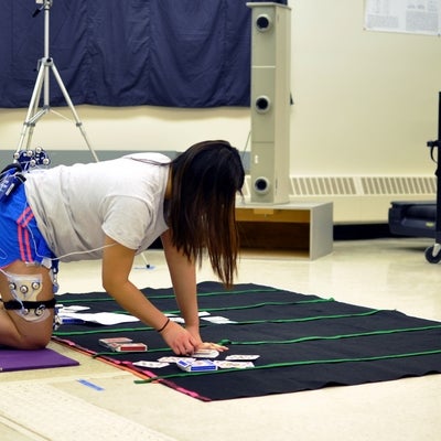
One of the high knee flexion postures measured by Professor Stacey Acker's students.

17 / 20
This subject is being exposed to 30 minutes of kneeling work to see if long periods of kneeling effects knee control mechanisms.

18 / 20
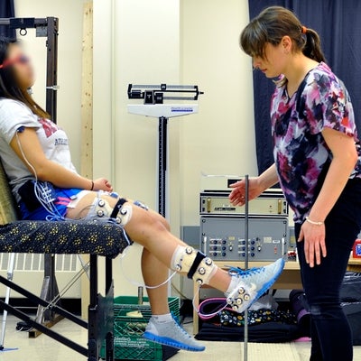
Preparing a participant for testing by fixing the shank on a nearly frictionless sled to ensure smooth controlled movements.

19 / 20
Instructing a blinded participant through a knee proprioception test to assess differences which may occur from prolonged kneeling work.

20 / 20
A participant performing a plantarflexed kneel while pressure is measured between the thigh and calf.
