Biomicro-electromechanical systems (BioMEMS) sensors

Nanofabrication

Nanofabrication is the process used to manufacture functional structures that are on the order of 100nm. Making components and structures at the nanoscale has many benefits, such as:

  • Unique physical, electrical, optical or magnetic properties not seen on the micro scale
  • Higher density
  • Lower cost
  • Increased performance

There are many ways to manufacture devices at the nanoscale, some of which are more conventional or "top-down", while others are more unique and follow the "bottom-up" approach. The Sensors and Integrated Microsystems Laboratory (SIMSLab) is actively researching in both top-down and bottom-up nanofabrication techniques.

Electron-beam lithography

Electron-beam lithography (EBL) is a "top-down" nanofabrication approach that uses electrons to expose an e-beam sensitive resist material, in a similar approach to conventional photolithography (Fig. 1). It is able to achieve very high resolution (10-20nm) and is extremely flexible, being able to pattern almost any geometry or layout desired (Fig. 2). However, EBL is a serial technique, making it slow and expensive. It also requires expensive equipment and highly controlled facilities, so it is generally used for research purposes and not manufacturing of commercial devices.

The SIMSLab has the capability to fabricate a variety of metal nanoparticle arrays using EBL at the facilities of the University of Western Ontario Nanofab and the  University of Toronto ECTI Nanofabrication. Such nanostructures exhibit unique optical properties (nanoplasmonics) that are being used in sensors that the SIMSLab is currently developing.

Nanosphere lithography

Nanosphere lithography (NSL) is a low cost nanofabrication technique used to produce triangular nanoparticles (Fig. 3). A monolayer of polymer microspheres, typically 400nm in diameter, self assembles on a clean substrate into a hexagonal close packed pattern (Fig. 4 and 5). It is then used as a mask for metal deposition; nanotriangles are formed between the interstitial sites of the crystal pattern. Removal of the microspheres leaves the nanotriangles array. Although lower cost, it is often difficult to maintain sufficient positional control, uniformity and repeatability with this technique (Fig. 6), which makes it difficult to use outside of research labs.

The SIMSLab has developed an in house NSL protocol to produce large areas of nanotriangles for nanoplasmonic sensor applications by using a spreading technique to produce the microsphere monolayer. However, it is still difficult to pattern nanoparticles in specific locations on the substrate. Therefore, they have also developed a new procedure called Geometrically confined NSL (GC NSL) (Fig. 7 and 8), which allows patterning of the nanoparticle arrays using conventional photolithography. This process reduces the cost from $250/mm2 with EBL to $0.10/mm2. This process is currently being developed for use in large scale manufacturing of low cost nanoparticle arrays for plasmonic sensing applications.

Label-free biosensors

SIMSLab is developing label-free, optical protein sensors for a variety of applications. Current methods for protein detection, such as enzyme-linked immunosorbent assay (ELISA) and the western blot, require trained personal, numerous reagents and equipment, and a large amount of time. Therefore, the SIMSLab is developing a portable protein sensing platform that can be used in the field for rapid protein detection by non-technical personnel.

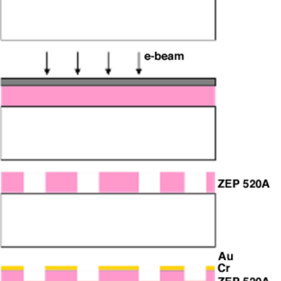
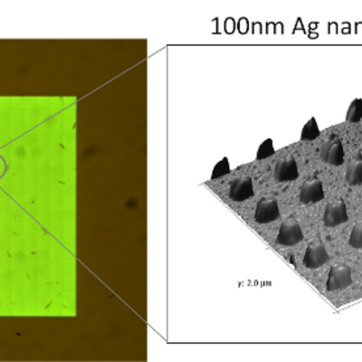
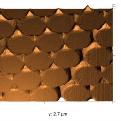
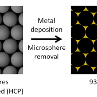
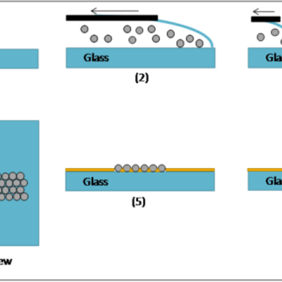
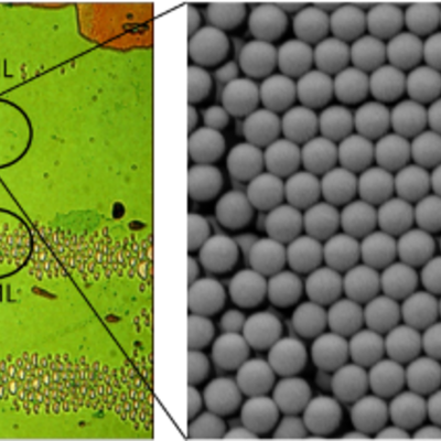
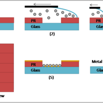
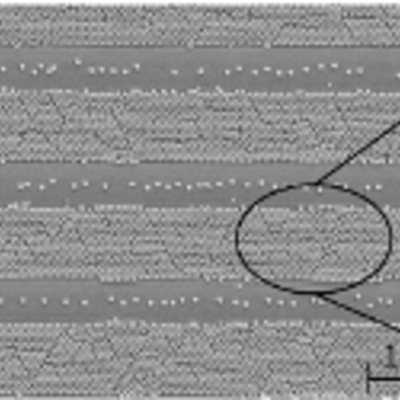
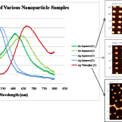
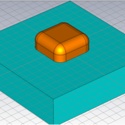
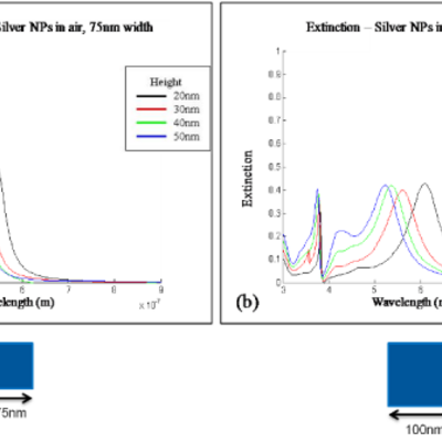
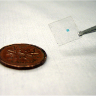
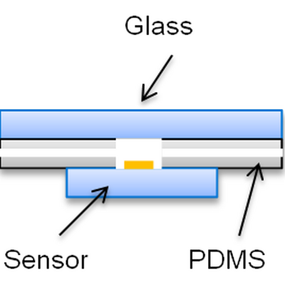
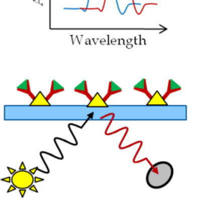
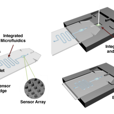
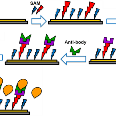
The protein sensing platform they have developed is based on localized surface plasmon resonance (LSPR) of metal nanoparticle arrays. Metal nanoparticle arrays are fabricated on glass using NSL or EBL (Fig. 13). Flow cells made of Polydimethylsiloxane (PDMS) are used along with a flow injection system to deliver samples to the nanoparticle array (Fig. 14). A simple white light source, spectrometer, and netbook are used to detect changes in the LSPR peak position upon protein binding (Fig. 15). The particle surfaces are functionalized with specific antibodies to capture only the proteins of interest. The prototype device is in a benchtop format, but is currently being developed into a portable system.

The advantages of the SIMSLab protein sensing platform are:

  • Label free technology
  • Low cost, batch fabrication sensors
  • Simple hardware requirements
  • Suitable for a wide variety of proteins and other biomolecules
  • Quantitative concentration readings with high specificity and sensitivity, and a low detection limit (ng/mL)
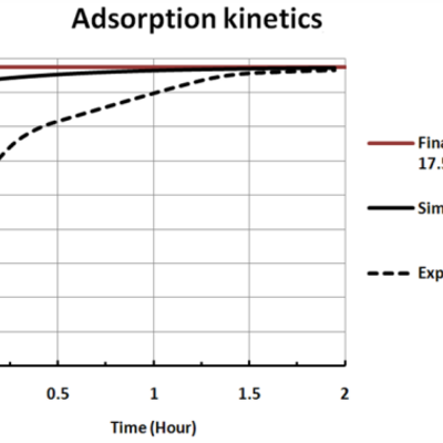
  • Measurement of protein kinetics
  • Minimal sample preparation
  • Robust against vibration, electromagnetic noise, and temperature fluctuations
  • Easily integrated into a handheld or portable device format
  • Fast response possible (less than 30 minutes)

Protein sensor for Heat shock protein 70 (HSP70)

HSP70 is a molecular chaperone protein, which performs multiple functions inside of cells, including protein translocation, stabilization and refolding of denatured proteins, higher order protein assembly, and degradation of irreversibly denatured proteins. HSP70 is an excellent marker of environmental health as it is highly conserved among species and upregulated in response to external stress stimuli, such as elevated environmental temperature, dehydration, osmotic stress, and heavy metal pollution. It also has the potential as a biomarker in health care, as high levels have been found in malignant tumours in breast, endometrial, bone, and gastric cancers. It is also a marker of early stage prostate cancer and cardiac health.

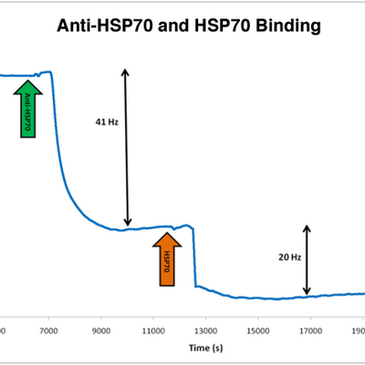
Through a collaboration with Matt Vijayan, an expert on HSP70, the SIMSLab has developed an LSPR-based protein sensor that is able to detect HSP70 without the use of labels. Selective capture of the protein is achieved using an anti-HSP70 antibody bound to functionalized metal nanoparticles. Shift in the LSPR peak position can be used to determine the concentration of HSP70 in a sample. We are currently optimizing the sensor to improve detection limits and develop a handheld version of the device.

Protein sensor for point-of-care medical diagnostics

Currently, the majority of medical labs cannot deliver blood work results within the ideal time, at which diagnosis is most critical and treatment most effective. For example, a bleed-to-read time of thirty minutes is ideal for diagnosis of myocardiac infarction (cardiac arrest). However, even the most advanced hospitals have difficulty obtaining results within one hour. Point-of-care (POC) diagnostics, in which the blood sample is analyzed at the patient bedside rather than being sent to the lab for analysis, offers rapid detection and at a lower cost. This increases hospital throughput, decreases spending, and increases patient diagnosis accuracy and treatment effectiveness.

The SIMSLab is developing multiplexed LSPR sensors for use in POC medical diagnostics (Fig. 16). Specifically, they are developing a sensor for simultaneous detection of four critical markers of myocardial infarction (MI): cTnT, cTnI, CK-MB, and Myoglobin. Detection of these biomarkers simultaenously offers a wealth of information regarding the type and severity of MI, as well as the potential for much earlier diagnosis. This sensor is currently under development and will be updated as progress is made.

Optimized surface functionalization

The realization of most BioMEMS sensors requires employment of a suitable functionalization protocol. Functionalization is defined as the formation of a bio-compatible interface on a transducing surface of a bio-chemical sensor, for immobilizing and subsequent sensing of biomolecules (Fig. 17). The SIMSLab has the capability to develop highly selective surface functionalization protocols for a wide range of proteins and small molecules.

SIMSLab has been developing functionalization protocols for immobilization of various antibodies and proteins such as Heat shock protein (HSP) 70 and biotin/streptavidin complex on planar and nanoparticle gold interfaces. Moreover, SIMSLab is working on in-situ functionalization procedures, which means performing the whole functionalization process inside of microfluidic channels. In-situ functionalization could greatly facilitate the production of portable BioMEMS sensors.

SIMSLab has also been enhancing the quality of the functionalization in terms of kinetics and receptor loading densities, which are critical parameters in performance and sensitivity of biosensors. The functionalization process involves attaching the protein of interest to an anti-body modified thiolic Self-assembled monolayer (SAM) formed on the gold substrates (Fig. 17). By modeling the SAM formation kinetics through the Finite element method (FEM) technique, SAM processing times in SIMSLab have been substantially reduced (Fig. 18 and 19). After FEM kinetic modeling, constitutive receptor conjugation parameters, such as crosslinking reagent concentrations, are carefully examined by Quartz crystal microbalance (QCM) (Fig. 20) and Atomic force microscopy (AFM) to determine the optimum protocol.

Current project members