Making it harder for cancer to hide
Health research at the University of Waterloo spans all Faculties. In systems design engineering, Alexander Wong and Hamid Tizhoosh are developing better imaging and scans for cancer treatment.

Health research at the University of Waterloo spans all Faculties. In systems design engineering, Alexander Wong and Hamid Tizhoosh are developing better imaging and scans for cancer treatment.
By Christian Aagaard Communications & Public AffairsAlexander Wong could have spun what he learned as a University of Waterloo student into a career in consumer electronics.
 Instead, he enlisted in the war on cancer.
An assistant professor in systems design engineering, Wong co-directs the Vision and Image Processing Lab with colleagues David Clausi and Paul Fieguth. Remote sensing, via satellite, is one side of his work. The other involves changing the way magnetic resonance imaging (MRI) works to reveal hard-to-find cancerous tissue.
“If we can pinpoint where the cancer is, it facilitates minimally invasive treatment,” Wong says. “We can destroy the cancerous region and leave the organ intact, with fewer side effects.”
Wong’s research currently focuses on prostate and skin cancers, conditions of particular concern to senior Canadians. Besides modulating MRI functions to “light up” cancerous tissue that is tricky to spot, his research group is developing accompanying clinical decision support software to improve how scans are read and interpreted by patient-care teams.
Wong expects clinical testing to begin next year at Sunnybrook Health Sciences Centre in Toronto.
“Our goal is to help doctors make consistent, well-informed decisions,’’ he says.
Cutting confusion
Hamid Tizhoosh, an associate professor in systems design engineering, focuses on quality assurance in cancer treatment. He and his students developed software to not only speed up the “segmentation” of medical scans, but also to improve the analysis that follows from it.
 Photo credit: Dan Epstein
Segmentation sorts an image into different regions, based on such things as intensity and texture. It’s a slow, labour-intensive process that, for example, helps distinguish diseased tissue from healthy tissue for diagnostic and treatment-planning purposes.
Trouble is, says Tizhoosh, different doctors will come up with different segmentations of the same scan.
But if that range of approaches could be collected and catalogued, “we could build a consensus contour, and hence, establish a baseline for accuracy measurement,’’ he says.
Through cloud computing and algorithms, his research group built “knowledge maps” — based on the input of many doctors, potentially from around the world — to achieve more consistent segmentations.
“This,” Tizhoosh says, “has the potential to practically eliminate the error in medical- image analysis, as the technology is based on collective wisdom of doctors.”

Read more
Waterloo prof leads a team of researchers to improve water quality through a community-focused approach underpinned by technical excellence

Read more
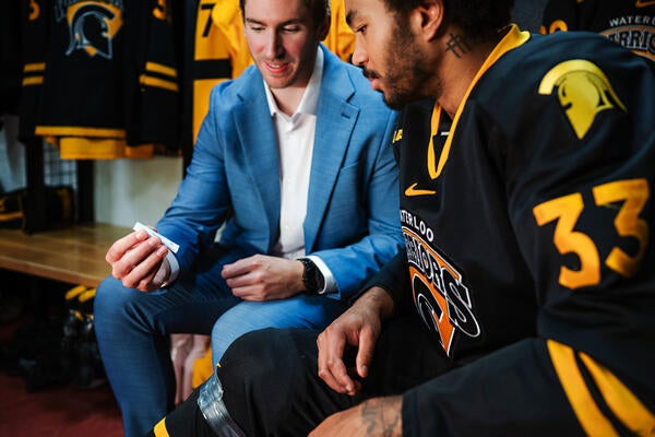
New medical device removes the guesswork from concussion screening in contact sports using only saliva

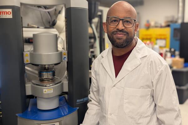
Waterloo researcher Dr. Tizazu Mekonnen stands next to a rheometer, which is used to test the flow properties of hydrogels. (University of Waterloo)
Read more
Plant-based material developed by Waterloo researchers absorbs like commercial plastics used in products like disposable diapers - but breaks down in months, not centuries
The University of Waterloo acknowledges that much of our work takes place on the traditional territory of the Neutral, Anishinaabeg, and Haudenosaunee peoples. Our main campus is situated on the Haldimand Tract, the land granted to the Six Nations that includes six miles on each side of the Grand River. Our active work toward reconciliation takes place across our campuses through research, learning, teaching, and community building, and is co-ordinated within the Office of Indigenous Relations.