Better diagnosing diseases with the help of AI
Researchers improve the trustworthiness of medical imaging diagnoses with innovative three-stage system powered by AI

Researchers improve the trustworthiness of medical imaging diagnoses with innovative three-stage system powered by AI
By Media RelationsAn interdisciplinary team of researchers at the University of Waterloo has developed a more trustworthy method to diagnose diseases such as COVID-19, pneumonia, and melanoma using artificial intelligence (AI) tools.
Waterloo engineering professor Alexander Wong teamed up with other researchers from the university, McGill University, and the National Research Council of Canada to develop a new method to support doctors by enhancing computer-aided diagnosis through deep learning.
The researchers created a system they’ve dubbed Trustworthy Deep Learning Framework for Medical Image Analysis (TRUDLMIA), which leverages the power of supervised and self-supervised AI learning that aims to pave the way for advancements in high-performing and trustworthy healthcare models.
“Models employing the TRUDLMIA approach outperform existing ones in various tasks, excelling in COVID-19, pneumonia, and melanoma diagnosis, surpassing models specifically designed for those tasks in terms of both performance and trust,” said project lead Dr. Wong, the Canada Research Chair in Medical Imaging and Artificial Intelligence. “The proposed system is undergoing further development to address future pandemics and syndromes, including long-term effects associated with COVID-19.”
Medical imaging and deep learning can improve medical AI by assisting with diagnosis, prediction and prognosis. To date, a key issue in using deep learning for medical image analysis has been the presence of data bias, low trust and accountability, and challenges in interpreting existing data.
TRUDLMIA’s innovation works in three stages to train the AI system. First, it learns from a very large set of labelled general data in a pre-training process, after which it learns from both general data and domain-specific data (e.g., medical data) without labels in a self-supervised learning process. Finally, it fine-tunes the AI with task-specific labelled data (e.g., clinical diagnosis data) with a special objective that focuses on handling data imbalances and biases and improving AI trustworthiness.
“TRUDLMIA, developed collaboratively with medical doctors, is poised to transform medical image analysis,” said collaborator Dr. Pengcheng Xi, Senior Research Officer at the National Research Council of Canada. “With a focus on improving diagnostic accuracy, boosting trust, and adapting across medical specializations, TRUDLMIA aspires to be an ally for doctors, to provide timely interventions for patients, and to support caregivers through preventive care.”
More information about this work can be found in the research paper, “Towards Building a Trustworthy Deep Learning Framework for Medical Image Analysis,” published recently in the journal Sensors.

Read more
New medical device removes the guesswork from concussion screening in contact sports using only saliva

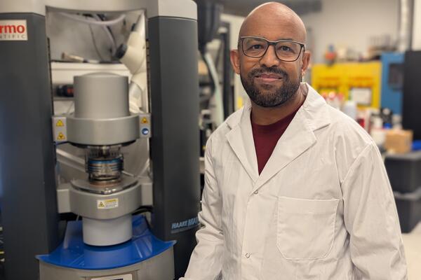
Waterloo researcher Dr. Tizazu Mekonnen stands next to a rheometer, which is used to test the flow properties of hydrogels. (University of Waterloo)
Read more
Plant-based material developed by Waterloo researchers absorbs like commercial plastics used in products like disposable diapers - but breaks down in months, not centuries

Read more
Here are the people and events behind some of this year’s most compelling Waterloo stories
The University of Waterloo acknowledges that much of our work takes place on the traditional territory of the Neutral, Anishinaabeg, and Haudenosaunee peoples. Our main campus is situated on the Haldimand Tract, the land granted to the Six Nations that includes six miles on each side of the Grand River. Our active work toward reconciliation takes place across our campuses through research, learning, teaching, and community building, and is co-ordinated within the Office of Indigenous Relations.