A retinal image could help doctors quickly distinguish between similar neurodegenerative diseases such as ALS and Alzheimer’s disease, and with remarkable accuracy, according to new research.
Researchers from the University of Waterloo are leading the development of a fast, non-invasive, and affordable diagnostic tool. There is currently no objective diagnostic test for ALS or Frontotemporal Lobular Dementia (FTLD-TDP), in which the protein TDP-43 forms deposits in the spinal cord and brain, respectively.
The new tool could help with earlier diagnosis, enabling timely interventions known to slow disease progression and supporting more targeted treatment development that could improve patient outcomes.
“This is a major step toward earlier and more accurate diagnosis,” said Dr. Melanie Campbell, professor emeritus of physics and optometry. “Right now, FTLD and ALS are diagnosed only after symptoms appear, which often means the disease is already advanced. Being able to detect these conditions earlier could transform how we treat them.”
The researchers used polarized light to image protein deposits in donated retinal samples from patients with Alzheimer’s and compared them to samples from patients with Frontotemporal Lobular Dementia with TDP-43 (FTLD-TDP) and Amyotrophic Lateral Sclerosis (ALS). By analyzing the light patterns, the team was able to accurately differentiate between the deposits of amyloid beta, associated with Alzheimer’s, from those with TDP-43, associated with FTLD and ALS, and predict the severity of the disease in the brain.
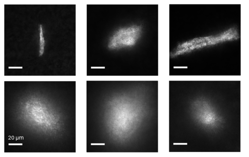
Samples of one of the 16 raw images taken in polarized light of retinal deposits. The left column shows sample deposits in an individual with brain pathology indicative of Alzheimer’s Disease; the middle column, pathology of ALS; the right column, pathology of FTLD (University of Waterloo).
They also uploaded data on the light interaction into two AI models, Random Forest, an ensemble learning method, and convolutional neural networks, an image-based method, to see if they could learn to differentiate amyloid beta from TDP-43 deposits. The differences between the two deposit types were strong enough that Random Forest predicted the right disease 86 per cent of the time, and convolutional neural networks were even higher at 96 per cent of the time.
“We hope that within a few years, this technology will evolve into a simple eye test capable of detecting and distinguishing multiple brain diseases, giving patients in smaller, underserved communities access to this type of testing,” Campbell said. “A fast, accessible diagnostic tool could make a profound difference for patients and families.”
The paper, Retinal Deposits of TDP-43 and Amyloid Beta and Associated Neurodegenerative Diseases are Accurately Classified using Measured Interactions with Polarized Light in Machine Learning Algorithms, appears in Alzheimer’s & Dementia: The Journal of the Alzheimer’s Association.