New laser imaging could precisely guide doctors during brain surgery
Researchers have taken an important step in the development of a microscope to precisely guide doctors during surgery to remove brain tumors

Researchers have taken an important step in the development of a microscope to precisely guide doctors during surgery to remove brain tumors
By Media RelationsResearchers have taken an important step in the development of a microscope to precisely guide doctors during surgery to remove brain tumors.
For the first time, a team led by engineers at the University of Waterloo used laser imaging technology to almost instantly identify cancerous tissue with accuracy comparable to laboratory tests that take up to two weeks.
That means the Photoacoustic Remote Sensing (PARS) imaging system could tell doctors where a tumor ends and healthy tissue begins so they know exactly where to cut.
“As you can imagine with the brain, doctors need to minimize the amount of tissue they remove because of the impact on the patient,” said Parsin Haji Reza, a systems design engineering professor who leads the project. “There is a very fine line.”

Two images, one produced in the lab (left) and the other using new PARS laser technology (right), show areas of cancer in brain tissue with comparable accuracy.
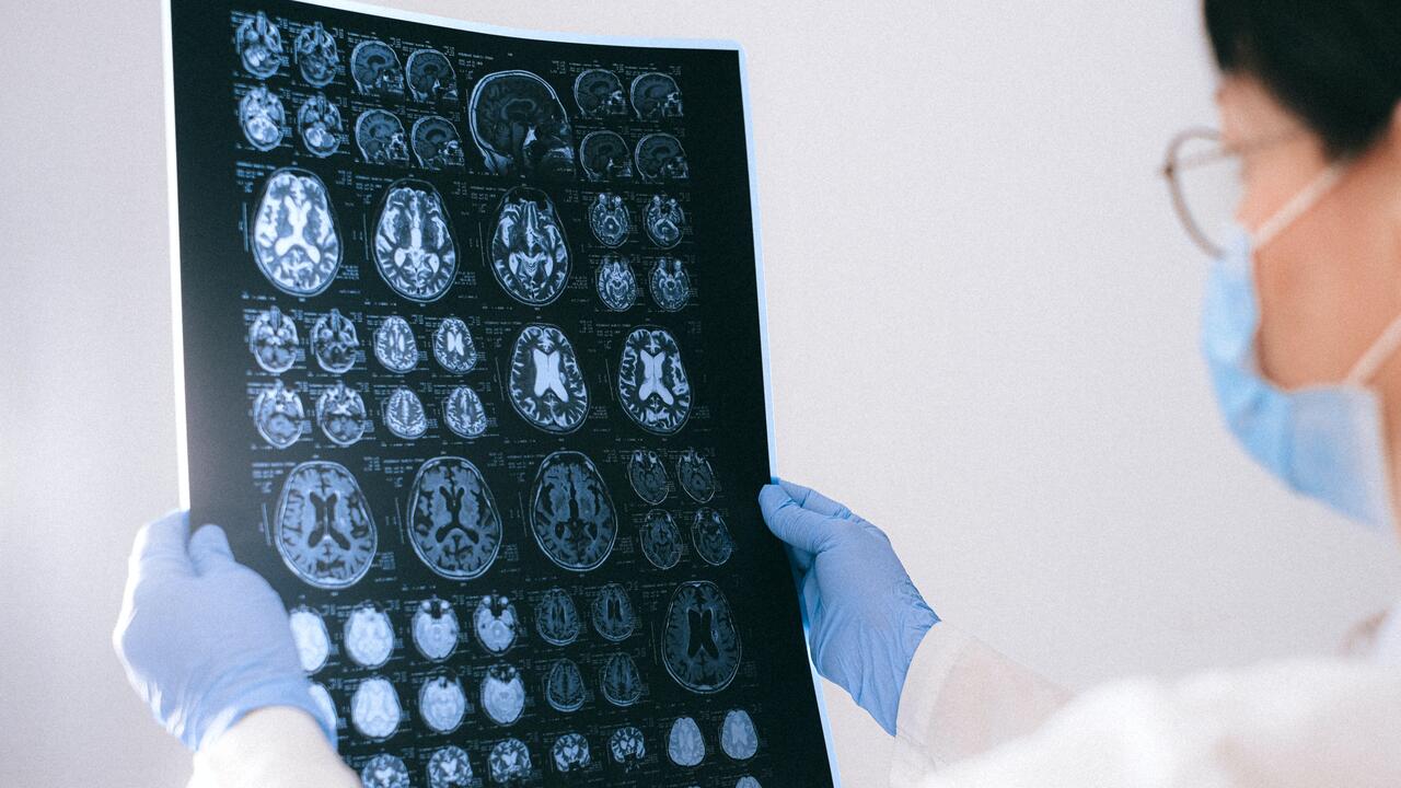
Doctors now rely on pre-operation CT scans and MRI images to locate brain tumors. Tissue samples are also examined during surgery, but tests take up to 30 minutes each and yield low-quality results.
To determine if they have entirely removed a tumor or not, surgeons must still send tissue samples to the lab for post-operative analysis that can take two weeks
A microscope developed by the researchers achieved results of similar quality to those ‘gold standard’ lab tests almost instantaneously.
“That opens up an extremely promising path towards our ultimate goal - a non-contact, surgical microscope that in real-time can guide doctors towards very safe, maximal resection with no waiting,” said Haji Reza, director of the PhotoMedicine Labs at Waterloo.
Missed malignant tissue during brain surgery is linked to poor clinical outcomes and lower survival rates.
The new technology works by sending multi-coloured laser pulses into tissue, which absorbs them, heats up, expands and produces sound waves. Another laser then reads the waves to determine if the tissue is healthy or cancerous
Researchers have started a company called illumiSonics to commercialize the system. They aim to have microscopes that quickly image tissue samples in operating rooms by the end of next year. Within three to five years, they hope to have developed a surgical microscope capable of imaging the brain itself during surgery without taking any tissue samples.
PARS technology has also been applied with promising results to breast, gastroenterological, skin and other kinds of cancer.
The research team includes graduate students Benjamin Ecclestone and Saad Abbasi, postdoctoral fellow Kevan Bell and engineering professor Paul Fieguth of Waterloo, and oncologists Deepak Dinakaran and John Mackey, and pathologist Frank van Landeghem of the University of Alberta.
A paper on their work, Improving Maximal Safe Brain Tumor Resection with Photoacoustic Remote Sensing Microscopy, appears in the journal Scientific Reports.

Read more
Robots the size of a soccer ball create new visual art by trailing light that represents the “emotional essence” of music

Read more
Waterloo prof leads a team of researchers to improve water quality through a community-focused approach underpinned by technical excellence

Read more
New medical device removes the guesswork from concussion screening in contact sports using only saliva
The University of Waterloo acknowledges that much of our work takes place on the traditional territory of the Neutral, Anishinaabeg, and Haudenosaunee peoples. Our main campus is situated on the Haldimand Tract, the land granted to the Six Nations that includes six miles on each side of the Grand River. Our active work toward reconciliation takes place across our campuses through research, learning, teaching, and community building, and is co-ordinated within the Office of Indigenous Relations.