AI-powered antenna revolutionizes bone fracture diagnosis
Waterloo Engineering researcher creates a new system to detect bone fractures that is fast, accurate and safe

Waterloo Engineering researcher creates a new system to detect bone fractures that is fast, accurate and safe
By Media RelationsA University of Waterloo engineer has paired inexpensive wireless communication antennas with artificial intelligence (AI) to improve how doctors can detect bone fractures.
Determining bone fractures using traditional diagnostic methods such as x-rays, computed tomography (CT) scans, and magnetic resonance imaging (MRI) takes time — such equipment is not readily available in ambulances or primary care facilities and, with health care services in high demand, many people have to wait for an x-ray or scan once they arrive at the hospital.
The new system delivers a faster, safer, more portable and cost-effective alternative to what currently exists.
“Our method is safer because it doesn’t expose patients to radiation or interfere with any medical devices in their bodies,” said lead researcher Dr. Omar Ramahi, a professor in Waterloo’s Department of Electrical and Computer Engineering.
“It’s also easy to transport, making it suitable for all people and situations, from elite athletes on the field to elderly nursing home residents to unconscious emergency room patients.”

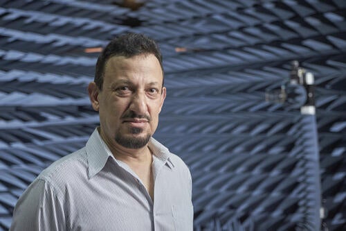
Dr. Omar Ramahi, a professor in Waterloo’s Department of Electrical and Computer Engineering and lead researcher for this study
Unlike conventional medical imaging methods that produce images requiring expert interpretation, Ramahi’s system detects cracks and breaks clearly, providing straightforward information that is crucial in emergencies.
It works by positioning two antennas on opposite sides of the suspected fracture site, with one antenna transmitting low-frequency microwaves through the bone to the other. The received data is then analyzed by a deep neural network — an AI model trained on extensive datasets of human body parts and bone fracture types.

An example of Dr. Omar Ramahi's lab set-up shows a cow leg bone sitting between a dipole antenna and a horn transmitting antenna. The horn transmitting antenna sends a microwave signal towards the bone which is then received and classified based on the type of fracture in the bone.
Ramahi’s system was developed in collaboration with an international research team and is the first to use AI with microwaves to detect bone fractures without using imaging techniques. Further work is underway to develop a high-resolution diagnostic tool. The researchers believe they could one day develop a portable device, potentially in the form of a cuff that wraps around the injured area, which could soon assist paramedics, long-term care staff and sports teams in immediate and preliminary fracture diagnosis.
“Our ambition is to advance this technology into a market-ready product,” said Ramahi. "We're eager to see this innovation reach its full potential to provide a rapid, inexpensive way to assess people’s injuries for bone fractures and speed up health care delivery.”
More information about this work can be found in the research paper, “Microwave bone fracture diagnosis using deep neural network”, published in IEEE 2023 53rd European Microwave Conference (EuMC).

Read more
New medical device removes the guesswork from concussion screening in contact sports using only saliva

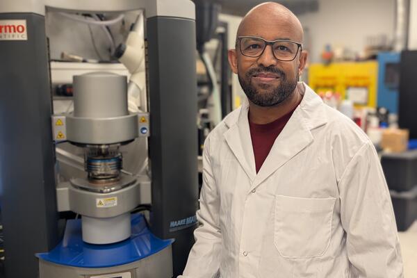
Waterloo researcher Dr. Tizazu Mekonnen stands next to a rheometer, which is used to test the flow properties of hydrogels. (University of Waterloo)
Read more
Plant-based material developed by Waterloo researchers absorbs like commercial plastics used in products like disposable diapers - but breaks down in months, not centuries

Read more
Here are the people and events behind some of this year’s most compelling Waterloo stories
The University of Waterloo acknowledges that much of our work takes place on the traditional territory of the Neutral, Anishinaabeg, and Haudenosaunee peoples. Our main campus is situated on the Haldimand Tract, the land granted to the Six Nations that includes six miles on each side of the Grand River. Our active work toward reconciliation takes place across our campuses through research, learning, teaching, and community building, and is co-ordinated within the Office of Indigenous Relations.