 Waterloo researchers in the Advanced Concepts Research Lab are developing a new method for medical imaging that could reduce the time to diagnosis.
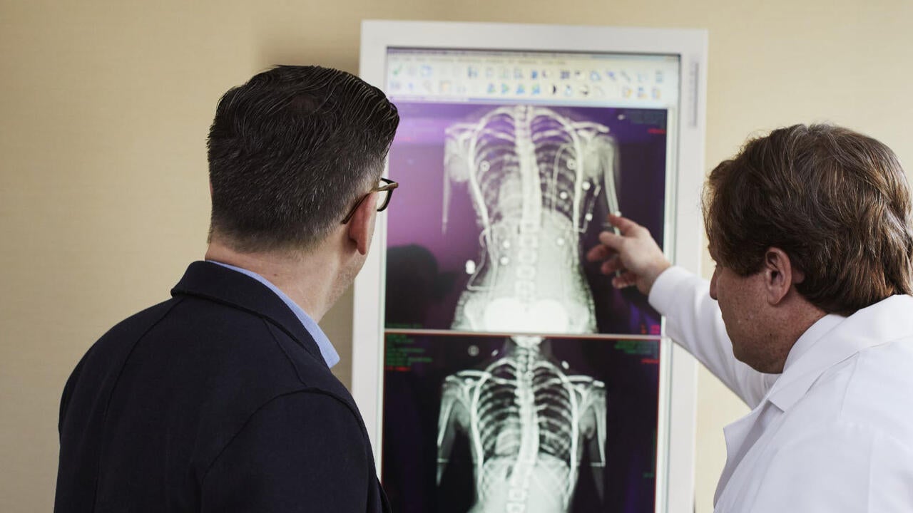
Clinicians currently rely on a number of technologies for medical imaging, including MRI, X-rays, and ultrasound. However, they all come with challenges. Safety, costly equipment, and low-resolution images can increase the time it takes to get a diagnosis, and patients can wait weeks before they’re able to begin treatment.
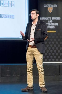
“A patient might get an X-ray, then come back in two weeks to get the results,” explains Seyed Hossein Mirjahanmardi, a doctoral candidate in electrical and computer engineering, and the principle researcher on the project. “Those two weeks can be crucial. If there was a device that was cheaper and safer, the doctor could do a test immediately in his office. We could get an initial image right away, and if the doctor sees something suspicious, he can send the patient for further examinations.”
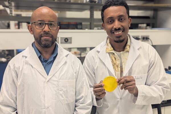
Supervised by professor Omar Ramahi, Mirjahanmardi and Ali Albishi, a collaborative researcher on the project, believe the solution lies in microwave technology. Using microwave imaging, they are creating a system that is safer way than other imaging technologies, like X-rays. Their system also relies on low-cost equipment, making it much cheaper than options like MRI.
Achieving a high resolution
Because microwave imaging uses a low frequency, the system is very safe to use. Unlike high-frequency X-ray imaging, microwave technology doesn’t involve any harmful radiation. Plus, it can easily penetrate through bodily materials that other options can’t, meaning that different layers of a body can be identified and included in a microwave image.
But traditionally, microwave technology produces a low-resolution image. This is the primary challenge Mirjahanmardi is trying to address.
“Microwave imaging has not been commercialized because of its low-frequency nature, which causes a low-resolution image,” he says. “Microwaves are very large, and they cannot detect small changes in the material that we are looking for.”
Mirjahanmardi uses simulations to mimic reality and create a clearer, accurate image. He begins with data from a transmitter-receiver system, and uses it in an algorithm that can identify small changes in a piece of tissue.
“Once we have collected the information from the receivers, we can use that data to properly track information and map it to a meaningful image,” he explains. “That image can show us where everything is – for example, where a tumour is in relation to other tissues.”
Reducing the time to diagnosis
Using a microwave device, a doctor could conduct a scan in his office – cutting out the waiting period necessary for more expensive and risky techniques.
“Our group is hoping to have the microwave image much sooner, taking the time frame from weeks to days,” Mirjahanmardi explains.
With his algorithm, the processing time could be significantly reduced. It’s possible that doctors and patients could receive the information in just a few days – helping them begin treatment as soon as possible.