January 22, 2016
Getting under your skin: A video camera that tracks blood flow
A Waterloo researcher talks about his passion for developing medical imaging technology that he hopes will save lives
A Waterloo researcher talks about his passion for developing medical imaging technology that he hopes will save lives
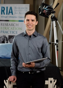
Robert Amelard, a University of Waterloo PhD student in systems design engineering, is making headlines with innovative technology that can help identify health problems before they result in a medical emergency. Waterloo Engineering sat down with Amelard to understand how the revolutionary technology works and what inspired him.
We’ve recently introduced a new portable touchless system that enables whole-body blood flow monitoring using patent-pending technology called Coded Hemodynamic Imaging (CHI). The system could lead to better detection and prevention of cardiovascular issues like heart attacks and strokes.
Imagine a world where we don’t rely on harmful events like a heart attack or stroke to realize that something is wrong. A technology gap exists and I saw the opportunity to propose a solution. I want to play an important role in the movement toward preventive healthcare by developing technology that provides for a more predictive, ongoing approach to monitoring health status. I hope to see this system used to identify early warning signs and really impact people’s quality of life.
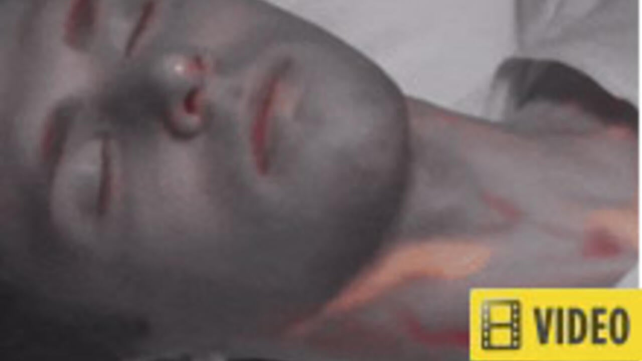
Accompanied by a light source, the device uses medical hardware design and digital signal processing to produce data and a visualization of blood pulsing at various parts of the body. More complex than what your typical video camera would capture, the device can see deeper into the skin, highlighting the blood flow. It’s safe - the device relies on the interaction of light with blood. No x-ray radiation or ultrasound, just the type of lights you would experience walking outside on a sunny day. We're actually looking for participants to validate the system so anyone who is interested can e-mail me at ramelard@uwaterloo.ca .
 For older adults this would enable ongoing assessment to identify blood flow problems. It could provide early warning signs of potential blockages. Where contact is an issue, the most vulnerable patients could benefit. Consider infants in neonatal intensive care, patients with painful burns or those with a highly contagious illness. And, with multi-person monitoring capabilities, this technology could have a major impact on emergency rooms and long-term care homes.
Anywhere contact is an issue or multiple persons require monitoring simultaneously, this device could be used to gain greater insights into health changes related to the vascular system. I want to see this device help as many people as possible.
A: Ultimately, my hope is to see it used in homes and doctor’s offices. In North America, health conditions are typically assessed under an acute healthcare model. In most cases treatment begins after you experience a physical symptom. Sometimes symptoms are reviewed in subjective ways that can vary from one physician to the next.
Getting this technology into the home and clinical environments could support a more preventive approach to managing individual healthcare needs by encouraging detailed health change monitoring.
Great advances are being made in healthcare, however, treatment is often only offered once a problem has been identified. The gap still exists between when something starts to go wrong in the body, and when it is diagnosed. Diagnosis usually happens after physical symptoms are experienced. And, after diagnosis, treatment can be delayed by lengthy hospital wait times. I want to help bridge the gap and develop solutions. There is so much we can learn from the body using imaging technology, which could result in better assessment and diagnostic tools.
Yes, medical applications for imaging technology have been an interest of mine since my undergraduate studies at Waterloo in software engineering. While completing my master’s degree, I developed a framework for image assessment to help doctors better analyze and diagnose potential cases of melanoma. With the support of my supervisors, Alexander Wong and David Clausi, I was able to identify my passion for developing medical imaging systems.