Background
Medical imaging is a vital aspect of modern health-care, with millions of images taken each year. Clinicians (cardiologists, urologists, oncologists etc.) and other health care professionals review these images in order to provide analysis and advice for diagnosis, treatment and palliation. It is a common task and activity in reviewing images to identify and delineate a segment of an image of interest. Such a segment may help to identify tumours, lesions, other regions of interest. It could also help pinpoint the location within the body of these regions of interest. Segmentation or contouring of a medical image is frequently necessary to provide patient diagnosis and prognosis, and to develop the patient care plan. Segmentation of images by radiologists is a laborious and time-consuming task which sometimes represents a significant bottleneck in providing timely patient treatment plans. Confounding this problem is the fact that significant radiologist interpretation variability can occur which can further impact patient outcomes.
Description of the invention
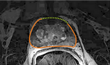
University of Waterloo (UW) researchers have developed a novel method for automatic image segmentation and auto-contouring which learns and improves its segmentation technique based on expert consensus segments (Atlas-based) as well as single user input.
Advantages
The advantages of this segmentation technique are as follows:
- Time/cost savings: faster, more consistent and more accurate segmentation saves user’s time, and improves patient outcome.
- Multi-modal: can be used for images derived from any imaging modality (eg. x-ray, ultrasound, etc.)
- Improved accuracy: expert consensus-based learning improves speed, accuracy and consistency across imaging modalities and platforms.
- Individualized learning: adapts to the preferences of each user, allowing consistent, individualized automatic segmentation.
- Efficient operation: fast retrieval of similar cases, leading to better diagnosis
Potential applications
Although this technology was developed with medical Imaging in mind, other potential applications include:
- Industrial robotics
- Autonomous vehicles
- Satellite image analysis
- Computer vision
Reference
7230 & 7312
Patent status
US 8391603B2
US 9361701B2
Stage of development
Working prototype
FDA approved for prostate image countouring
Ongoing research
Contact
Scott Inwood
Director of Commercialization
Waterloo Commercialization Office
519-888-4567, ext. 33728
sinwood@uwaterloo.ca
uwaterloo.ca/research