Design team members: Niles Guo, Siwan Kim, Candy Wong, Steve Zuo
Supervisors: Professor John Yeow and Albert Chen
Background
A typical configuration of an ultrasound catheter system consists of three basic components: an ultrasound generator, an ultrasound transducer, and an ultrasound catheter. An ultrasound transmission member, such as a wire, transmits vibrational energy from an ultrasound transducer to a distal head of the catheter to disrupt blockages in blood vessels, also known as vascular occlusions.
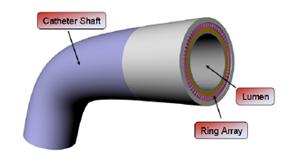
Figure 1: CMUT Ring Array Catheter Technology [1]
Intracardiac imaging (IVUS), defined as imaging from within the cardiac chambers and the major blood vessels, differs from intravascular ultrasonographic imaging (ICE) that refers to ultrasonographic navigation and visualization in small blood vessels (usually coronary arteries). Both procedures make use of similar catheter designs, however, a key difference is that ICE requires more steering and maneuverability.
Minimally invasive catheter-based interventional electrophysiology (EP) is becoming a standard in the management of cardiac arrhythmias. The use of non-fluoroscopic ICE catheters is a better solution for guiding EP procedures, since it improves procedural efficiency and reduces the undesirable use of fluoroscopy, which is currently the common catheter guidance method.
Project description
The main objective of this project is to create a miniaturized ultrasound catheter system. An ultrasound catheter system is a type of medical imaging that uses high frequency broadband sound waves that are reflected by tissue to produce images. The main application of this technology is for imaging cardiovascular tissues in order to assess potential damage and illness.
Currently, the ultrasound catheter system is used in two types of sonographies, Intravascular Ultrasound (IVUS) and Intracardiac Echocardiography (ICE). Two-dimensional ICE is the primary echo modality used in today's electrophysiology laboratory because it provides meaningful, real-time feedback on the catheters positioned within the anatomy of the heart. However, due to its size it poses many limitations to its applications such as maneuverability and ease of use. Therefore, it is advantageous to develop a miniaturized version of the existing technology to improve its ease of use and lower cost.
Design methodology
The project is sectioned into three distinct phases: Research, Design/Build and Testing. Within each phase, there will be iterations of work to ensure objectives are achieved before moving onto the next phase.
Research Phase
- Build and assembly of current CMUT device to gain firsthand experience of the assembly and integration processes the unit
- Investigation for decreasing the size of the device
- Investigation on improving image quality
Design/Construction Phase
- The most appropriate research result would be used that takes account of the cost, practicality, and appropriate technical level
- Designs will be compared with the objectives and the most appropriate one will be chosen
- Once a final design is decided, the group would move into the build phase, and work to create the working prototype
Testing Phase
- The device will be tested to see if it meets all of the objectives set forth at the beginning of the project
- If there is an aspect of the prototype that did not meet the objective, the research results will be utilized to see if there are other alternatives available
- Material testing for the final prototype to ensure overall quality
References
Nikoozadeh, Amin. Ömer Oralkan, Mustafa Gencel, Jung Woo Choe, Douglas N. Stephens, Alan de la Rama, Peter Chen, Kai Thomenius, Aaron Dentinger, Douglas Wildes, Kalyanam Shivkumar, Aman Mahajan, Matthew O’Donnell, David Sahn and Pierre T. Khuri-Yakub. "Forward-Looking Volumetric Intracardiac Imaging Using a Fully Integrated CMUT Ring Array." Print.