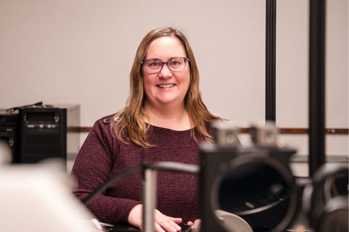
Dr. Jennifer Hunter develops advanced fluorescence imaging techniques to examine individual cell layers, which could lead to earlier retinal disease detection.
By Karen Kawawada
Fluorescence isn’t just for highlighters, lighting or clothes that look trippy at a black light party. For Dr. Jennifer Hunter, an associate professor at the University of Waterloo School of Optometry and Vision Science, fluorescence is the key that will unlock ways of detecting diseases of the eye and brain earlier and help test treatments for these diseases.
Hunter, who joined the University of Waterloo in 2022, holds a Discovery Grant from the Natural Sciences and Engineering Research Council of Canada and two U.S. National Institutes of Health research grants for projects started when she was a faculty member at the University of Rochester. She was named an Association for Research in Vision and Ophthalmology Silver Fellow last year.
Before she got into fluorescence, though, Hunter thought she might go into medicine, so she enrolled in biology at the University of Waterloo. Quickly realizing her real interests lay in physics, particularly optics and imaging, she changed her major.
Hunter completed her undergraduate degree, followed by a master’s and then a PhD, both supervised by Dr. Melanie Campbell. After her joint physics-vision science doctorate, she headed to the University of Rochester in 2006 for a postdoctoral fellowship.
A snag in research plans
Hunter joined the lab of Dr. David Williams, a pioneer of adaptive optics. This field allows for clear imaging of cells in the living eye by correcting for natural optical imperfections using methods similar to the ones astronomers use on high-powered telescopes to adjust for the presence of the Earth’s atmosphere.
For her postdoc, Hunter was supposed to lead a project using imaging to better understand how children’s retinas develop after birth. Soon after she arrived, however, a graduate student discovered that the imaging techniques being used were causing unexpected changes to the retinas of animal models. As a precaution, all human ocular imaging was shut down.
A new focus on light safety
Rather than dwelling on her ruined project, Hunter turned her focus to learning more about both the retinal changes that had been observed and about light and laser safety.
The retina has multiple layers of cells, of which the retinal pigment epithelium (RPE) can be thought of as the caretaker of the retina. RPE cells recycle and transport molecules, and in doing so, they accumulate byproducts over time that become a conglomerate called lipofuscin.
Lipofuscin is highly fluorescent, which is useful in imaging. The normal pattern of lipofuscin was disrupted in animal models by the light used in imaging studies at Rochester.
“It turned out that existing light safety standards were not adequate to protect the retina against changes that we were able to observe with our cellular-scale imaging methods,” says Hunter.
Hunter now sits on the American National Standards Institute (ANSI) Z136 Accredited Standards Committee and the Laser Bioeffects and Medical Surveillance subcommittee. She helped write the ANSI Safe Use of Lasers standards that set laser use safety guidelines for everything from medical use to industrial applications to laser shows.

Dr. Jennifer Hunter (centre) works with Michelle Peimann, research manager (left), and Dr. Rosa Martínez Ojeda in her lab.
Detecting early changes in cells
Using fluorescence to study light safety inspired Hunter to use it to learn more about the retina.
“What I'm really interested in is how we can detect early changes in the function of retinal cells,” says Hunter. “The literature has reports of people missing half their photoreceptors and still having 20/20 vision, so by the time you have a patient complaining of vision loss, they may be at the stage where a lot of cells are dead or irreversibly damaged. We want to detect changes long before that point.”
Hunter’s work on improving retinal imaging could help diagnose patients with very early-stage diseases and monitor how patients respond to treatment.
“In cases of retinal degeneration, if a patient finds that they’ve lost a number of lines on the visual acuity chart, they’ve gotten to the point of retinal cell death or complete non-function,” says Hunter. “If you can use imaging to measure the function of retinal cells before they die, the assessment is a better measure than the visual acuity chart. Using imaging, we can potentially test whether a drug designed to target a specific process in a cell is working properly. That’s the big-picture motivation for the work I do.”
Measuring nanoseconds of light
Hunter’s Advanced Ophthalmoscopy Waterloo Laboratory is working on further refining cellular-scale fluorescence imaging, including aspects such as the intensity of fluorescence, the exact colour being emitted, and the time delay in emission, because these are factors that can change with disease.
Her lab has developed an adaptive optics fluorescence lifetime imaging ophthalmoscope (FLIO), which enables unique visualisation of individual layers of cells in the living eye. In addition, Hunter’s lab has a clinically styled FLIO system made by Heidelberg Engineering – one of only about a dozen in the world and the only one in Canada.
FLIO measures how long certain molecules in the eye glow after being hit by laser light. This very short duration, called a lifetime, is measured in fractions of nanoseconds – one-billionth of a second. Lifetimes of molecules vary according to many factors, including the type of molecule and environmental factors such as temperature and pH. There’s strong evidence that lifetimes get longer in diseased cells.
Hunter’s lab uses both standard single-photon excited fluorescence and two-photon excited fluorescence. Two-photon excited fluorescence means that instead of using one shorter-wavelength photon, molecules are hit almost simultaneously by two longer-wavelength photons. Excited molecules briefly emit light. Using two photons instead of one allows imaging of molecules that couldn’t otherwise be seen. Hunter’s work was the first to successfully carry out two-photon excited fluorescence imaging through the pupil of the living eye.
“A lot of people thought two-photon fluorescence imaging of the living eye would be impossible because of light safety issues,” says Hunter. “They said similar things about FLIO. I’m really proud of having achieved something other people were skeptical about.”
Better imaging could lead to treatments
One of Hunter’s current projects focuses on imaging the retinas of people with Stargardt disease, a progressive genetic disease that leads to vision loss at a young age. Since the disease is thought to relate to an accumulation of lipofuscin within RPE cells, better imaging of this cell layer could lead to the development of treatments.
Hunter’s imaging techniques could be applied to diseases involving other cell layers and molecules too. One of her proudest accomplishments has been separately imaging transparent layers of the retina that have historically been hard to image. This includes the ganglion cell layer, which is affected in glaucoma.
“There’s lots of suggestion now that Alzheimer’s disease, diabetes and other systemic diseases can be visible in changes in the retina at a very early stage,” says Hunter. “My goal is to image the retina better so that we can observe what changes might be occurring with normal function, aging and disease. From there, we can try to develop new treatments to prevent blindness.”
Dr. Jennifer Hunter is currently recruiting graduate students and postdoctoral fellows. For more information, see Vision Science Graduate Studies.