Design team members: Benjamin Alton, Sabrina Cannistraro
Supervisor: Dr. Ed Jernigan
Project description
The team will develop and implement an algorithm that allows for accurate and efficient diagnosis of chest interval changes in children from sequential digital x-rays. This will be accomplished through the use of pre-processing image enhancement techniques, image alignment, digital subtraction, and post-processing image enhancement techniques.
Background
Digital images are becoming increasingly popular in the field of medicine. The Hospital for Sick Children has recently transformed their medical imaging medium from film to digital images and has reduced the time between taking an x-ray and providing it to the doctor with a signed report by 70% [1].
Digital images are also becoming widely used in Computer Aided Diagnosis (CAD) of various diseases including cancer, pneumonia, and lung nodules among others. CAD is currently in its infancy but slowly beginning to become more prominent for diagnosis in adults. One CAD algorithm that has clearly demonstrated its benefit in a number of studies involves the subtraction of two digital images. The resulting difference image shows nodules and tumours more clearly than the original images.
It is clear that the need for this type of diagnostic tool for children is no less dire and the benefits that such a system could add would be no less striking.
Design methodology
The general design methodology used for this project involves five steps of design. First, pre-processing of images will be performed to enhance image detail. Image registration will then be performed, which consists of two steps: Determining the relative position of the two images, and interpolating to find and apply an appropriate transformation that matches the images. Then, digital subtraction of the images will be performed to produce a single difference image as output. Finally, image processing techniques will be performed on this resulting image to enhance details of the image. These steps are illustrated in the system diagram in Figure 1.
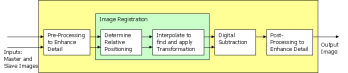
Figure 1: System diagram
Each of these five steps is treated independently, each including its own design alternatives. Each step is evaluated based upon the extent to which it meets the design goals: Accuracy of results and speed of processing. The best design from each step will then be incorporated into one final design. Iterative tests are performed on the final design against the overall design goals, and improvements are made as necessary.
Algorithm design
Preliminary image processing
The images being compared in this project can be of very low contrast meaning often detail is difficult to distinguish. The preliminary image processing may or may not be a useful tool to initially enhance the images. The team’s first task was to identify potentially useful preliminary image processing techniques, and evaluate their effectiveness to aid in image registration. Two preliminary image processing techniques were investigated. Though good results were achieved, the team decided against preliminary image processing because it sacrificed processing time.
Control point matching
The first step necessary in the alignment or registration of images is to determine the position of the images with respect to each other. One method often used for image registration is the use of control points. That is, a number of distinctive points, representative of the shape and form of a first image, are “matched” to points in the second image which are the most similar. An algorithm is implemented to help find the “best matches” for these control points in the other image. The image on which control points are originally selected is often called the “master”. Similarly, the image for which the best match points are found is called the “slave”.
Figure 2 below depicts graphically the algorithm used by the team to match control points.
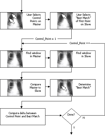
Figure 2: High level control point matching algorithm
Results
The images in Figure 3 through 6 depict the master and slave image used to complete the testing. The control points on the master are visible as X’s and the “best matches” as determined by the algorithm are similarly visible on the slave image.
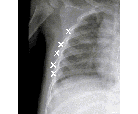
Figure 3: Master image with control points (left side)
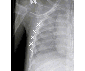
Figure 4: Slave image with "best match" points determined by optimal algorithm (Left Side)
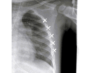
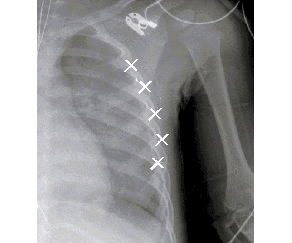
Figure 6: Slave image with "best match" points determined by optimal algorithm (Right Side)
The team has designed and implemented the first two steps in their algorithm. The remainder of the project, as described below, is completed as part of SYDE 462 in January to April 2002.
Interpolate to find and apply transformation
The team's next step for this project is to interpolate between the control points found, and transform the slave image such that it aligns with the master image. This allows constant portions of the two images to be cancelled when subtraction is performed in the next step, to give an output image which only illustrates the difference between the subsequent images.
Digital subtraction
In this section of the project, the team will subtract the transformed slave image from the master image to reveal differences between the two subsequent images.
Post-processing to enhance detail
Finally, the team will once again investigate different image enhancement techniques to improve the appearance of the difference image to aid in more accurate and efficient diagnosis.
Reference
[1] GE Medical Systems, Press Release, December 21, 2000.