Grant recipients:
Anton Trinh, Caryl Russell, and Robert Burns, Kinesiology
(Project timeline: May 1, 2019 - April 30, 2020)
Project team:
Anton Trinh, Caryl Russell, Robert Burns, Tamara Maciel, and Sarah Mitchell-Ewart, Kinesiology
Description
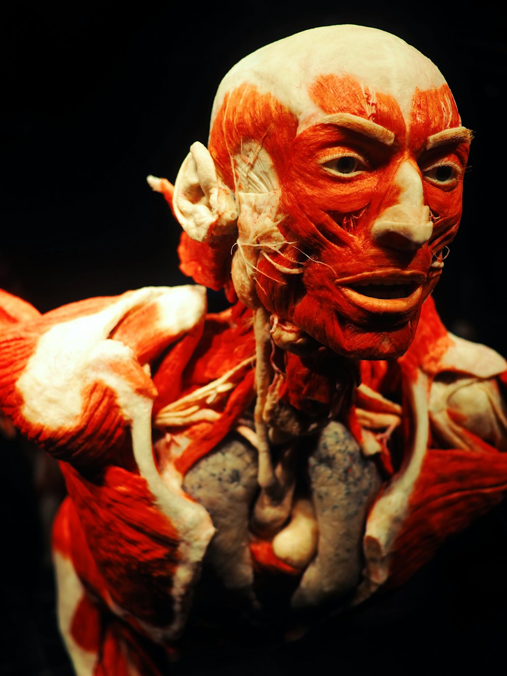
Students in Kinesiology require an understanding of surface anatomy (external features of the human body) in relation to the internal structures, such as muscles or nerves. However, students have difficulty linking what they palpate or see superficially to the anatomy below the skin. Instructors have indicated that this is due to both a knowledge gap and limited useful resources available to review and practice.
This project aims to fill this knowledge gap. The primary objectives are 1) to provide 3D anatomical modelling software (AMS) on wireless devices used in lab and 2) create related video content from the software. Videos will be made available within LEARN for unlimited access. These resources will contribute to a blended learning model that will facilitate student understanding between external features and internal structures, and improve hands-on skills resulting from deeper anatomical understanding. Student learning outcomes and resource use will be assessed.
Questions Investigated
Overall, this project assessed whether 3D resources improved learning outcomes requiring working knowledge of internal and external anatomical structures applied to hands-on skills such as palpation. Additionally, insight into student resource preferences was examined.
To investigate whether improvements occurred, grades (in aggregate) were compared between this project’s cohort (2019) and prior cohorts. In addition, instructor perception of students’ working knowledge was surveyed. To assess student preferences between 2D and 3D resources, user statistics from LEARN were examined. Specifically, “number of students accessing” a resource and “time spent viewing” were analyzed.
Findings
Overall, 3D resources did not have an impact on student performance as assessed by practical evaluation marks or overall grades between cohorts provided with 3D resources and those without. However, instructors and students did indicate a preference for 3D resources. Survey results indicated 3D resources were preferred over 2D resources by both instructors and students.
Students self-perceived to have stronger working knowledge of anatomical structures whereas instructors did not perceive students to have improved working knowledge (with respect to previous cohorts). Students recalling 3D resources, reported greater confidence in their working knowledge and subsequent hands-on skills.
References
Azer, S. A., & Azer, S. (2016). 3D anatomy models and impact on learning: A review of the quality of the literature Elsevier BV. doi:10.1016/j.hpe.2016.05.002.
Miller, M. (2016). Use of computer‐aided holographic models improves performance in a cadaver dissection‐based course in gross anatomy. Clinical Anatomy, 29(7), 917-924. doi:10.1002/ca.22766.
Peterson, D. C., & Mlynarczyk, G. S. A. (2016). Analysis of traditional versus threedimensional augmented curriculum on anatomical learning outcome measures. Anatomical Sciences Education, 9(6), 529-536. doi:10.1002/ase.1612.
Pringle, Z., & Rea, P. (2018). Do digital technologies enhance anatomical education? Retrieved from https://www.openaire.eu/search/publication?articleId=core_ac_uk__::ab946b08a3667a27 29bd15e9b50eea16.
Wilkinson, K., & Barter, P. (2016). Do mobile learning devices enhance learning in higher education anatomy classrooms? Retrieved from http://eprints.mdx.ac.uk/17589.